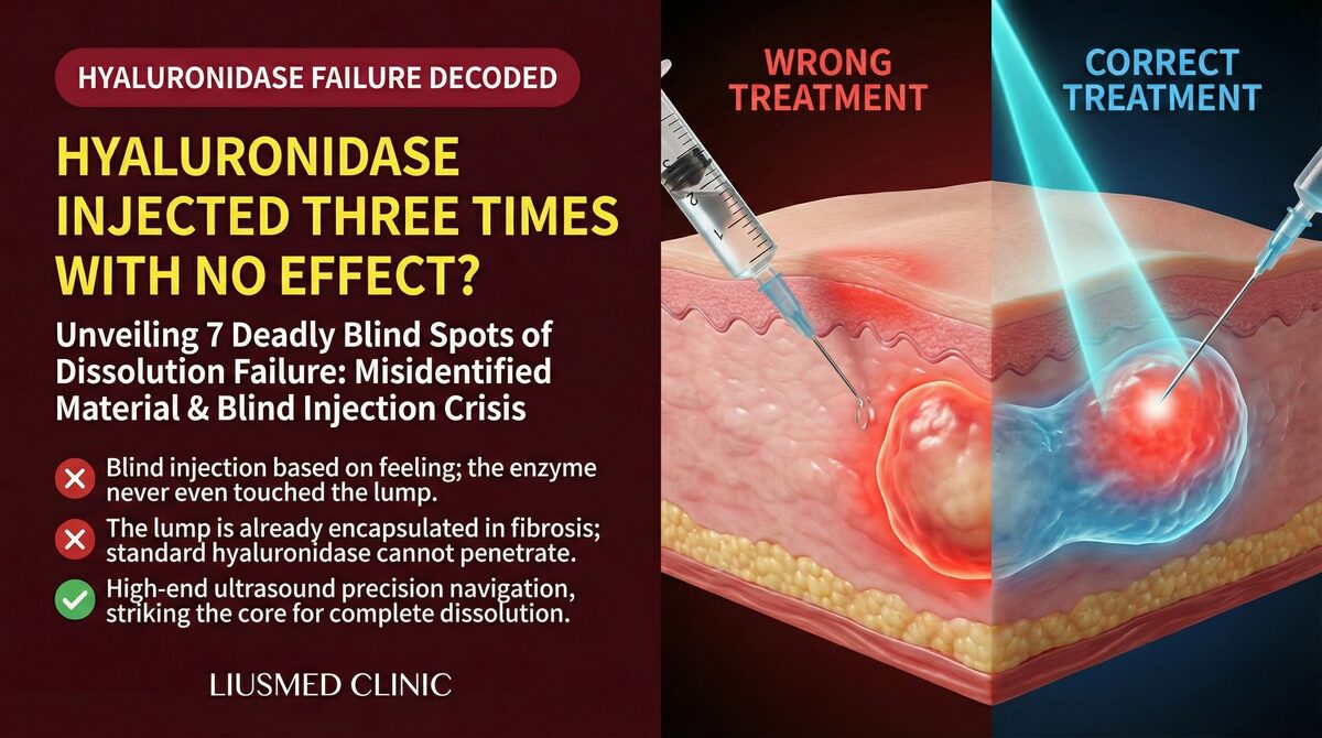
"I've had hyaluronidase injected three times and the lump is still there. What now?"
"It's a collagen stimulator — there's no enzyme that can dissolve it. But I don't want surgery either."
These are among the most common questions in my clinic. Many patients assume "ultrasound-guided" means "using ultrasound to enhance dissolving." In reality, ultrasound is an imaging navigation tool — entirely separate from the strategic choice between dissolving and extracting.
This article breaks down both dimensions: dissolution (chemical strategy) vs extraction (physical strategy), and the role ultrasound plays in each. For a broader overview of revision approaches, see Treatment Methods Overview.
Physical Extraction vs Chemical Dissolution: The Fundamental Difference
Start with where the failed material has gone and what condition it is in.
How Chemical Dissolution Works
Hyaluronidase cleaves the glycosidic bonds of hyaluronic acid polymer chains, breaking HA molecules into smaller fragments the body can metabolize. This mechanism works well for unencapsulated, correctly positioned, genuinely HA filler.
But for the "won't-dissolve lumps" seen in practice, failure clusters around these causes:
← Swipe to see more →
| Failure Reason | Explanation |
|---|---|
| Encapsulation | The body has walled the filler in a dense fibrous capsule the enzyme cannot penetrate |
| Non-HA material | Collagen stimulators (PCL, PLLA, CaHA), silicone, PAAG — no corresponding enzyme exists |
| Biofilm contamination | Bacterial biofilm attached to filler surface; even if the gel dissolves, inflammation persists |
| Migration | Filler has drifted to unintended tissue planes; enzyme injection site is inaccurate |
Each round of hyaluronidase also dissolves the body's natural HA — a major component of the skin matrix; the enzyme is not selective. Cumulative use may lead to skin thinning and tissue laxity, though how much thinning follows how many rounds has not been reliably quantified, and animal work has reported single-dose hyaluronidase without significant structural change. The judgement isn't the round count — it's that material which won't dissolve shouldn't keep being dissolved. For the mechanism in detail, see The Cumulative Damage of Repeated Hyaluronidase.
How Physical Extraction Works
Physical extraction bypasses chemical reactions entirely. It creates a direct entry point and removes the material and capsule tissue together.
The essential prerequisite: you must know what you're removing and where it is. Blind aspiration (no image guidance) has a documented history of vascular and nerve injuries. By contrast, ultrasound-guided physical extraction lets the physician confirm before any instrument enters:
- The exact location and depth of the filler
- Whether encapsulation is present, and how thick and adherent the capsule is
- The course of adjacent vessels and nerves
- The echo characteristics of the material (HA remnant, calcified CaHA, PLLA granuloma)
"You cannot safely treat what you cannot see" — this is why physical extraction can replace chemical dissolution in the right scenarios.
Key Insight: Ultrasound here is the "eyes," not the "weapon." It does not accelerate dissolution or emit energy to break down filler. It gives the operator spatial awareness throughout the procedure — enabling precise entry and safe clearance.
Hyaluronidase vs Physical Extraction: Choosing by Clinical Scenario
There is no universal answer to "which is better." The decision rests on three intersecting variables: material type × encapsulation grade × patient expectations.
Material-Based Triage
← Swipe to see more →
| Material | Hyaluronidase Applicable? | Physical Extraction Feasible? |
|---|---|---|
| HA (hyaluronic acid) — unencapsulated | ✅ First choice, highly efficient | Feasible but excessive |
| HA — encapsulated / repeated failure | ⚠️ Poor penetration, damages native HA | ✅ Recommended |
| HA — migrated / Tyndall effect | ⚠️ Dissolution may worsen asymmetry | ✅ Precise extraction more predictable |
| PCL (Ellansé) | ❌ No corresponding enzyme | ✅ Primary option |
| PLLA (Sculptra) | ❌ No corresponding enzyme | ✅ Granuloma extraction |
| CaHA (Radiesse) | ❌ No corresponding enzyme | ✅ Pinhole extraction feasible |
| Permanent fillers (silicone, PAAG) | ❌ No corresponding enzyme | ✅ Sequential extraction strategy |
Encapsulation Grade Determines Priority
Grade 0–1 (soft nodule): Hyaluronidase still effective, especially for HA nodules within 3–6 months of injection.
Grade 2–3 (palpable firm lump): Capsule has formed; enzyme penetration is weak. Marginal return on further chemical dissolution diminishes while each round damages native HA. Physical extraction should be considered.
Grade 4 (stone-hard, calcified): Calcified CaHA nodules or long-standing PLLA granulomas — hyaluronidase is completely ineffective. Extraction is the only viable approach.
Patient Expectations vs Recovery Time
Chemical dissolution has a shorter recovery window but may require multiple sessions with uncertain outcomes. Physical extraction is typically one procedure with a defined clearance scope, but carries a short recovery period (mild swelling 1–2 weeks depending on extent).
Key Insight: The "let's try hyaluronidase first and see" approach for encapsulated lumps creates a negative cycle — each failed round depletes native HA and accelerates tissue atrophy, making subsequent physical extraction technically more difficult. When ultrasound confirms encapsulation, proceeding directly to physical extraction is the more efficient path.
Filler Removal Method Comparison: Open Surgery, Blind Aspiration, Ultrasound-Guided
Many patients realize only during consultation that "filler removal" is not synonymous with "surgery." Three technical pathways exist clinically:
Path A — Open Surgery (Incision and Removal)
Indicated for extensive material involvement, severe migration, or large-scale adhesions. Larger wound with scar management required. Advantage: complete visualization, maximum material clearance.
Path B — Blind Aspiration / Blind Curettage
Historically, some clinics aspirated filler with needles or cannulas without image guidance. Literature documents vascular injuries, nerve damage, and post-operative depressions from blind aspiration. A pre-ultrasound fallback approach — not recommended as standard practice today.
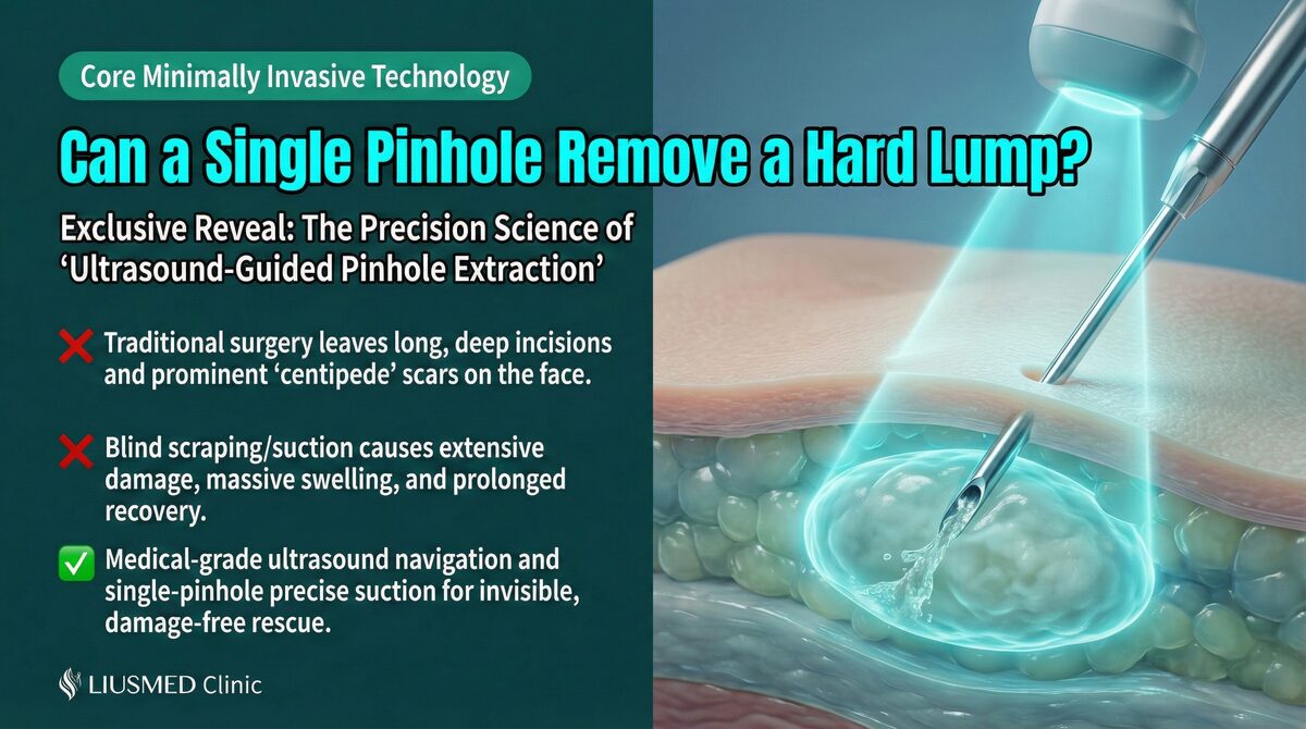
Path C — Ultrasound-Guided Single-Pinhole Physical Extraction
The minimally invasive technique Dr. Liu advocates. Key steps:
- Pre-operative ultrasound scan: Confirm 3-D filler location, echo type (HA / PCL / calcified / granuloma), capsule thickness, adjacent vessels
- Single-entry design: Select the shortest-path, vascular-free pinhole (approximately 1mm incision)
- Real-time ultrasound guidance: Operate under continuous screen monitoring — see instrument tip, see material movement, see clearance confirmed
- Material confirmation: Extracted tissue visually confirms material type, informing post-procedure assessment
Single-pinhole vs multi-portal: Multiple portals theoretically cover more area but each additional entry adds infection pathway and scar risk. Single-pinhole requires more precise localization but minimizes total trauma.
← Swipe to see more →
| Dimension | Open Surgery | Blind Aspiration | Ultrasound-Guided Single-Pinhole |
|---|---|---|---|
| Visualization | Complete | None | Real-time ultrasound image |
| Trauma | Larger | Small (but blind injury risk) | Minimized, predictable |
| Safety | High (requires anesthesia) | Low (blind) | High (guided throughout) |
| Indication | Extensive severe adhesion | Not recommended | Most localized firm lumps |
| Scar | Present | Minimal but possible depression | Minimal (≈1mm) |
For further clinical detail on the single-pinhole technique, see Filler Lump Extraction Technique Explained.
Ultrasound-Guided Extraction Principles: You Cannot Safely Treat What You Cannot See
Ultrasound guidance is relatively new in aesthetic revision medicine — worth explaining in depth.
What Ultrasound Reveals
Cutaneous filler characteristics on ultrasound imaging:
- HA (hyaluronic acid): Hypoechoic (anechoic), usually well-defined borders; encapsulated forms show hyperechoic fibrous ring
- CaHA (Radiesse): Hyperechoic with acoustic shadowing; calcifications are conspicuous
- PLLA (Sculptra): Granules appear uniformly hypoechoic; granuloma phase shows hyperechoic core
- PCL (Ellansé): Uniformly hypoechoic, similar to HA; history and palpation assist differentiation
- Vessels: Color Doppler mode shows blood flow; mandatory pre-procedure scan
Dynamic Intraoperative Monitoring
Static localization is just the start. During the procedure, the operator confirms:
- Whether the instrument tip is at the capsule margin or inside the filler mass
- Whether the filler is fully cleared or residual material remains (residual volume determines whether a second portal is needed)
- Whether adjacent microvasculature has been disturbed (any abnormal echo changes)
This real-time dynamic monitoring is the advantage ultrasound guidance holds over static MRI — while MRI has superior resolution, it cannot be used intraoperatively in real time.
Why Not Every Clinic Offers Ultrasound-Guided Extraction
Ultrasound operation requires specialized training (the operator must be fluent in soft-tissue ultrasound imaging), higher equipment cost (high-resolution linear probe), and longer operating time. These factors leave many clinics relying on "palpation + judgment + hyaluronidase" or advising patients to wait for natural absorption.
For patients, choosing a physician with ultrasound guidance capability means: more accurate pre-operative assessment → fewer unnecessary hyaluronidase rounds → lower intraoperative vascular risk.
Non-Dissolvable Filler Removal Options: PCL, CaHA, and Silicone
This section addresses the area of greatest patient confusion: what to do when a "permanent" or "long-lasting" filler develops a complication.
PCL (Ellansé) Nodules
PCL (polycaprolactone) is biodegradable but very slowly (labeled longevity: S/1yr, M/2yr, L/3–4yr). Nodules typically appear 6–18 months post-injection as fibroproliferative nodules.
Options:
- Watchful waiting (nodule may shrink as PCL degrades, but timeline is unpredictable)
- Ultrasound-guided physical extraction — for palpable PCL nodules, single-pinhole can directly remove the polymer core
- Intralesional corticosteroid injection (short-term reduction, not curative, skin atrophy risk)
For material comparison context, see HA Filler Revision vs Simple Dissolution.
CaHA (Radiesse) Calcified Nodules
In some patients, calcium hydroxylapatite particles calcify further, appearing as strongly hyperechoic on ultrasound with stone-like palpation. Hyaluronidase is ineffective.
Options:
- Ultrasound-guided physical extraction: acoustic shadowing precisely locates the calcified focus; single-pinhole can clear small-to-medium calcified nodules
- Open surgery: for extensive calcification or deep-plane involvement
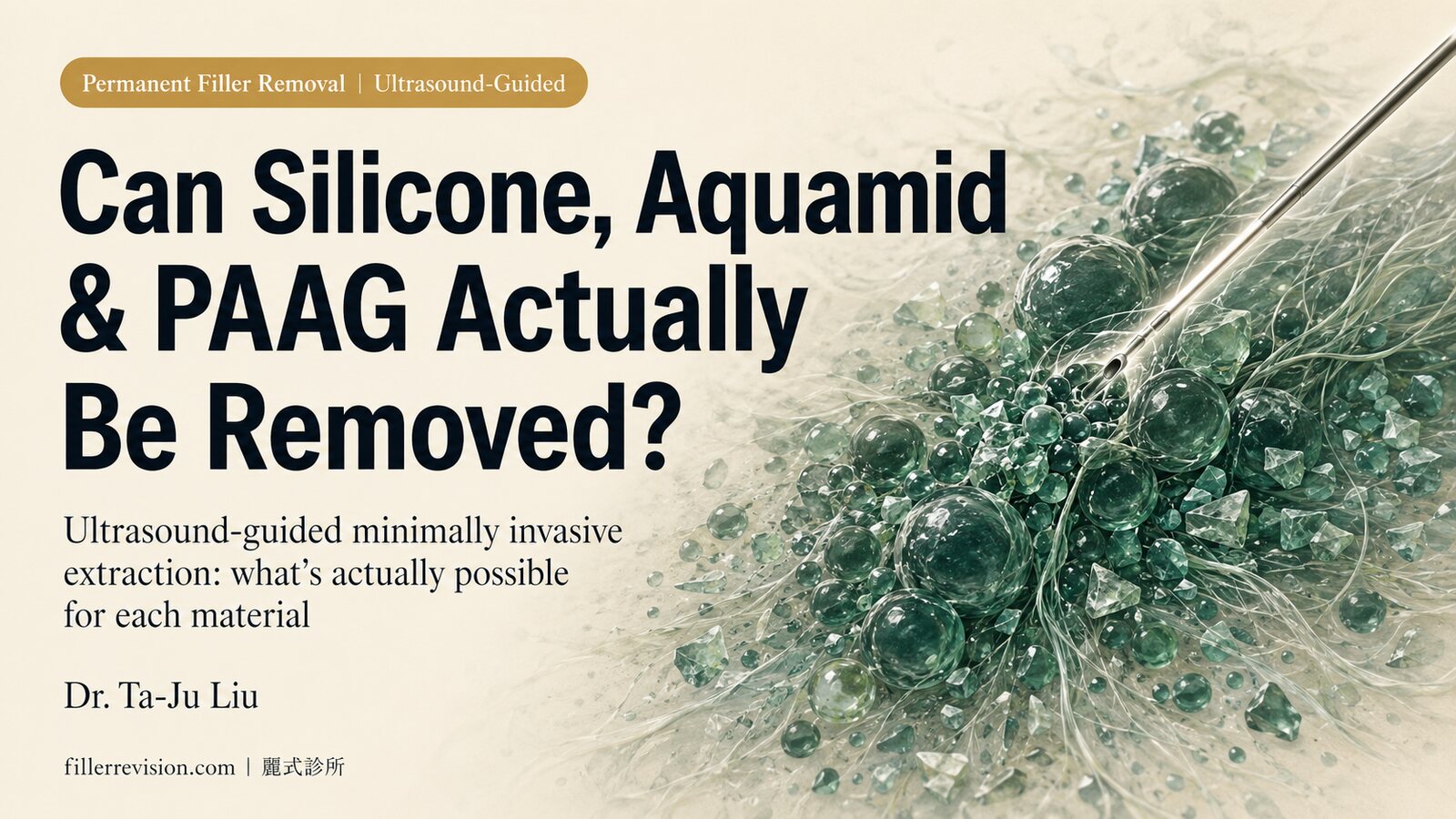
PAAG / Silicone and Other Permanent Fillers
Polyacrylamide hydrogel (PAAG, also known as "Amazingel" or "Bioformacryl") and liquid silicone were common permanent fillers in the 2000s, now banned in Taiwan and mainland China, but patients who received earlier injections continue to present.
Extraction challenges:
- Liquid silicone is highly infiltrative, permeating tissue spaces without clear borders
- PAAG disperses as multiple small foci in tissue; ultrasound shows diffuse hypoechoic deposits
- "Complete removal" cannot be guaranteed — the goal is "substantially reduce residual burden" and "clear the symptomatic primary foci"
For full analysis, see Permanent Filler Removal: Silicone, Aquamid, and PAAG.
FAQ
Q1. My filler is HA. Hyaluronidase has been injected twice and the lump remains. Third round or switch to physical extraction?
I recommend ultrasound evaluation before a third round. If imaging confirms encapsulation (hyperechoic fibrous ring surrounding the lump), further hyaluronidase will have limited effect and each round continues to deplete native HA. For encapsulated HA lumps, ultrasound-guided physical extraction is the more direct resolution.
Q2. How large is the single-pinhole incision, and how long is the recovery?
The portal is approximately 1mm — typically requiring a single small stitch or no suture. Post-operative swelling depends on extent of clearance, usually subsiding within 5–10 days. Most patients do not need time off work, though exercise and irritating cosmetics should be avoided for 1–2 weeks. Individual recovery varies; a medical evaluation establishes realistic expectations.
Q3. Is ultrasound-guided physical extraction appropriate for any type of filler lump?
It is most effective for well-defined, localized lumps — including encapsulated HA nodules, PCL fibroproliferative nodules, small-to-medium CaHA calcifications, and localized PLLA granulomas. Widely infiltrative permanent fillers (such as liquid silicone) cannot be cleared in a single session and require a staged extraction strategy.
Q4. I've seen claims online that HIFU can "dissolve" filler. Is that the same as ultrasound-guided extraction?
Completely different. High-intensity focused ultrasound (HIFU, as in "Ultherapy") uses thermal energy to tighten subcutaneous tissue — it does not target filler material. There are clinical case reports of HIFU accelerating filler migration or triggering inflammation in patients with existing filler. "Ultrasound-guided" in the context of extraction refers to diagnostic imaging mode — a low-energy linear probe displays real-time structural images to guide instrument movement. No heating, no tissue energy delivery.
Summary and Next Steps
The choice between physical extraction and chemical dissolution is not about "which is better." It depends on:
- Whether the material can be dissolved (collagen stimulators and permanent fillers have no corresponding enzyme)
- Degree of encapsulation (capsule thickness determines enzyme penetration rate)
- Number of prior failed attempts (each hyaluronidase round has a cost)
- Your expectations and recovery tolerance (one-time clearance vs repeated attempts)
If you have had two or more rounds of hyaluronidase with persistent lumps, or the material is confirmed to be a collagen stimulator or permanent filler, consider booking a consultation with Dr. Ta-Ju Liu. Bring any records of prior injections and ultrasound images if available.
You may also review the Filler Revision Service page for the full scope of what is offered before deciding.
Filler revision is precision medicine, not a cycle of repeated attempts. You cannot safely treat what you cannot see.
Written by Dr. Ta-Ju Liu. Reviewed under the Filler Revision Clinic editorial policy. For educational purposes only — does not replace individual medical evaluation.