What Happens When Your Body Builds a Wall Around Your Filler?
If you have tried dissolvers — perhaps multiple rounds — and the lump is still there, you are probably wondering why the treatment that was supposed to work simply did not. The answer lies in a biological process that many patients are never told about: encapsulation. Your body has built a collagen wall around the filler, and no amount of enzyme injected outside that wall can reach the material sealed inside.
This process, called encapsulation, is one of the most significant yet least discussed complications in aesthetic medicine. It is the primary reason why enzymatic dissolvers—including hyaluronidase for HA (Hyaluronic Acid) fillers—sometimes fail completely, leaving patients with persistent lumps that no amount of medication can resolve.
The Biology of Capsule Formation
Stage 1: The Acute Inflammatory Response
Within hours of injection, the body launches an acute inflammatory response. Immune cells—primarily macrophages and neutrophils—migrate to the injection site to investigate the foreign material. These cells release inflammatory mediators that increase blood flow to the area, causing the redness, swelling, and tenderness commonly experienced after filler injections.
In most cases, this acute inflammation resolves within days as the immune system determines that the filler material is biologically inert and does not pose an active threat. However, the immune system does not simply forget that the material is there.
Stage 2: Chronic Foreign Body Response
After the acute phase subsides, the body transitions to a chronic foreign body response. Macrophages that cannot fully digest the filler material fuse together to form multinucleated giant cells—large, specialized cells that attempt to engulf and break down foreign objects. When the filler material is too large or chemically resistant to be digested, these giant cells persist at the filler surface indefinitely.
Think of this like a group of workers trying to move a boulder. Individually, they cannot move it. So they surround it and attempt to contain it instead.
Key insight: At FILLER REVISION, our clinical experience confirms that the foreign body response is not a sign that something has gone wrong — it is the body's normal, expected reaction to any implanted material. The question is not whether this response will occur, but how severe it becomes and whether it leads to clinically significant encapsulation that blocks all non-surgical treatment options.
Stage 3: Fibroblast Recruitment and Collagen Deposition
As the chronic foreign body response continues, the body shifts its strategy from attempting to digest the filler to containing it. Fibroblasts—the cells responsible for producing structural proteins—are recruited to the filler surface. These fibroblasts begin depositing collagen fibers in organized layers around the filler deposit.
Over weeks to months, these collagen layers accumulate to form a distinct capsule wall. The capsule is composed primarily of Type I and Type III collagen, arranged in concentric layers that progressively thicken and densify over time.
Stage 4: Capsule Maturation
As the capsule matures, it undergoes structural changes that make it increasingly impermeable:
- Collagen cross-linking: The collagen fibers within the capsule become chemically cross-linked, increasing the structural rigidity and reducing permeability
- Vascular regression: Blood vessels within and around the capsule become sparse, reducing the delivery of immune cells and therapeutic agents to the filler surface
- Myofibroblast contraction: Specialized contractile cells within the capsule wall can cause the capsule to tighten around the filler, creating a firm, palpable nodule
The result is a dense, avascular, chemically cross-linked collagen barrier that effectively seals the filler off from the surrounding tissue environment.
Why Dissolvers Cannot Penetrate the Capsule
The Barrier Effect
Enzymatic dissolvers such as hyaluronidase are large protein molecules. To dissolve filler material, these enzymes must physically contact the HA chains and cleave specific chemical bonds. When a mature fibrous capsule surrounds the filler, the enzyme faces an impenetrable barrier.
Consider the analogy of trying to dissolve sugar inside a sealed glass jar by pouring water over the outside of the jar. No matter how much water you use, the sugar inside the jar remains untouched because the glass prevents the water from reaching it. The fibrous capsule functions in exactly this way—it prevents the enzyme from reaching the filler material it is designed to dissolve.
Key insight: The failure of dissolvers against encapsulated filler is not a matter of dosage or technique. It is a fundamental physical limitation: the enzyme cannot pass through the capsule wall, regardless of how much is injected into the surrounding tissue.
What Actually Happens When You Inject Dissolvers Near an Encapsulated Filler
When hyaluronidase is injected into tissue containing an encapsulated HA filler, the following occurs:
- The enzyme diffuses through the surrounding tissue but is blocked by the capsule wall
- Native HA in the surrounding tissue is degraded, which can cause localized volume loss, skin thinning, and textural changes in the area around the capsule
- The encapsulated filler remains intact because the enzyme never reaches it
- The patient experiences tissue damage from the enzyme without any benefit to the encapsulated filler
This creates a particularly frustrating clinical scenario: the dissolver causes visible damage to the healthy tissue surrounding the capsule while having zero effect on the problem material inside.
Factors That Increase Encapsulation Risk
Not all fillers are equally likely to become encapsulated. Several factors influence the likelihood and severity of capsule formation:
← Swipe to see more →
| Factor | Higher Risk | Lower Risk |
|---|---|---|
| Filler type | Non-biodegradable, silicone, PMMA (Polymethyl Methacrylate) | HA (early treatment) |
| Injection volume | Large bolus deposits | Small, distributed volumes |
| Injection depth | Incorrect tissue plane | Appropriate anatomical depth |
| Duration in tissue | Years | Months |
| Repeated injections | Multiple sessions, same site | Single treatment |
| Patient biology | Strong foreign body responders | Mild responders |
| Biofilm presence | Contaminated filler | Sterile filler |
How to Identify Encapsulated Filler
Clinical Signs
Encapsulated filler typically presents as:
- A firm, well-defined lump or nodule that is palpable under the skin
- A mass that does not change in size or consistency over time
- A deposit that does not respond to massage, compression, or dissolving agents
- A lesion that may or may not be visible, depending on its depth and location
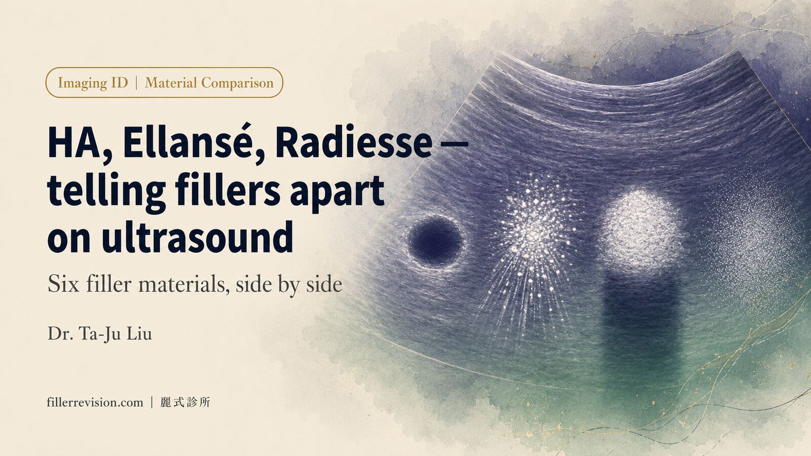
Ultrasound (Ultrasonography) Characteristics
High-frequency ultrasound is the most reliable imaging modality for identifying encapsulated filler. Characteristic findings include:
- Hyperechoic rim: The fibrous capsule appears as a bright, well-defined border surrounding the filler deposit
- Defined margins: Unlike non-encapsulated filler, which has irregular or blending borders, encapsulated filler has sharp, clear boundaries
- Internal heterogeneity: The filler material inside the capsule may show mixed echogenicity, reflecting partial degradation, fragmentation, or inflammatory changes
- Acoustic shadowing: Dense, calcified capsules may produce posterior acoustic shadows similar to those seen with calcified structures
Key insight: Ultrasound assessment before attempting dissolution is essential. If imaging reveals encapsulation, proceeding with hyaluronidase alone will be ineffective and may cause unnecessary tissue damage.
What This Means for FILLER REVISION Patients
Encapsulation is one of the most common reasons patients arrive at FILLER REVISION after failed dissolver treatments elsewhere. Understanding this biology explains why those treatments did not work — and why the path forward is different. At FILLER REVISION, we use high-resolution ultrasound to confirm whether encapsulation has occurred before recommending any treatment. If imaging reveals a capsule, we know that enzymatic dissolution alone will be ineffective, and we can proceed directly to a strategy designed to address the capsule barrier. This diagnostic-first approach prevents unnecessary tissue damage from repeated dissolver injections and leads to more predictable, lasting results.
The Primary Solution: Physical Extraction Through the Capsule Wall
Why Extraction Works
Physical extraction succeeds where dissolvers fail because it addresses the fundamental problem: the capsule itself. By physically accessing the interior of the capsule, the physician can remove the filler material regardless of whether it is chemically dissolvable.
The extraction process involves:
- Ultrasound-guided localization: Precisely identifying the capsule location, dimensions, and relationship to surrounding structures
- Targeted access: Creating a controlled entry point through the capsule wall using appropriate instrumentation
- Material removal: Extracting the filler material from within the capsule through aspiration, curettage, or a combination technique
- Capsule management: Depending on the clinical situation, the capsule wall itself may be partially removed, collapsed, or left to remodel naturally
- Verification: Real-time ultrasound confirmation that the material has been successfully removed
What About the Capsule Itself?
After filler extraction, the empty capsule typically undergoes one of two outcomes:
- Natural remodeling: In most cases, the capsule wall softens and is gradually remodeled by the body's normal tissue turnover processes. The dense collagen is broken down and replaced with normal tissue over weeks to months.
- Persistent capsule: In some cases, particularly with thick, mature capsules, the capsule wall may persist as a palpable but soft residual structure. If clinically significant, additional intervention may be needed to address the capsule itself.
The Classification of Capsule Severity
Not all capsules are equally challenging. A grading system helps guide treatment decisions:
Grade I — Mild encapsulation: Thin capsule, minimal fibrosis, filler partially accessible to dissolvers. Hyaluronidase may achieve partial success. Combined approach (enzyme + extraction) is often effective.
Grade II — Moderate encapsulation: Well-formed capsule with significant collagen deposition. Dissolvers achieve minimal penetration. Physical extraction is recommended as primary treatment.
Grade III — Severe encapsulation: Thick, dense, possibly calcified capsule. Dissolvers are completely ineffective. Physical extraction with capsule management is required.
Grade IV — Complex encapsulation: Multiple encapsulated deposits, anatomically challenging location, or associated complications (biofilm, granuloma). Comprehensive extraction strategy with ultrasound guidance is essential.
Can Encapsulation Be Prevented?
While encapsulation cannot be entirely prevented—it is a fundamental biological response to foreign materials—several strategies can reduce its likelihood and severity:
- Use biodegradable fillers when possible. Materials that the body can partially or fully degrade are less likely to trigger severe encapsulation.
- Avoid large bolus injections. Distributing volume in smaller deposits reduces the foreign body surface area and the intensity of the immune response.
- Inject at the correct tissue depth. Material placed in anatomically appropriate planes integrates more naturally with surrounding tissue.
- Limit repeated injections to the same site. Cumulative volume and repeated trauma increase encapsulation risk.
- Address complications early. If you notice persistent firmness or lumps developing, early evaluation and intervention can prevent progression to severe encapsulation.
Failed Dissolvers Do Not Mean No Solution — FILLER REVISION Can Help
If you have been told that your filler cannot be dissolved, or if multiple dissolver treatments have failed, the problem is likely encapsulation — not an untreatable condition. At FILLER REVISION, Dr. Ta-Ju Liu specializes in ultrasound-guided extraction of encapsulated filler material, providing a definitive solution when enzymatic approaches have reached their limit. Understanding why dissolvers failed is the first step toward a treatment that actually works.
Frequently Asked Questions
I have done several rounds of dissolver injections, but the lump is still there. Why didn't it work?
When dissolvers fail repeatedly, the most likely reason is encapsulation: your body has built a collagen wall around the filler that the enzyme cannot pass through. Hyaluronidase has to physically contact the filler to work, but a mature fibrous capsule blocks it like a sealed glass jar keeps water away from the sugar inside. This is a physical limitation, not a matter of dose or technique, so injecting more enzyme into the surrounding tissue does not reach the sealed-off material.
Is it harmful to keep injecting dissolver into an encapsulated filler?
Yes, it can cause harm without any benefit. When hyaluronidase is injected near an encapsulated filler, the enzyme is blocked by the capsule wall and never reaches the filler, so the lump stays intact. Meanwhile the native HA in the surrounding healthy tissue is degraded, which can cause localized volume loss, skin thinning, and textural changes. The result is visible damage to the healthy tissue while the problem material inside the capsule is unaffected.
How can I tell if my filler is encapsulated before trying another dissolver treatment?
High-frequency ultrasound is the most reliable way to identify encapsulated filler. On imaging, a capsule shows a bright, well-defined hyperechoic rim and sharp, clear margins around the deposit, unlike non-encapsulated filler which has irregular or blending borders. Clinically, encapsulated filler tends to be a firm, well-defined lump that does not change over time and does not respond to massage or dissolving agents. Assessing with ultrasound before attempting dissolution helps avoid an ineffective treatment that may cause unnecessary tissue damage.
If dissolvers can't work, what is the solution for encapsulated filler?
The primary solution is physical extraction through the capsule wall. It succeeds where dissolvers fail because it addresses the capsule itself: by physically accessing the interior, the filler can be removed regardless of whether it is chemically dissolvable. The process uses ultrasound-guided localization, a controlled entry point through the capsule wall, removal of the material by aspiration or curettage, and real-time ultrasound confirmation that it has been removed. At FILLER REVISION, Dr. Ta-Ju Liu uses ultrasound-guided extraction of encapsulated filler when enzymatic approaches have reached their limit.
After the filler is extracted, does the empty capsule stay in my face?
In most cases the empty capsule does not stay. After extraction, the capsule wall typically softens and is gradually remodeled by the body's normal tissue turnover, with the dense collagen broken down and replaced by normal tissue over weeks to months. In some cases, particularly with thick, mature capsules, the wall may persist as a palpable but soft residual structure, and if it is clinically significant, additional intervention may be needed to address the capsule itself.
Can encapsulation be prevented, and what raises the risk?
Encapsulation cannot be entirely prevented because it is a fundamental biological response to any implanted material, but its likelihood and severity can be reduced. Large bolus injections and a long duration of filler in the tissue both increase the risk, as do repeated injections to the same site and the use of non-biodegradable materials. Helpful strategies include using biodegradable fillers when possible, avoiding large bolus injections by distributing smaller volumes, injecting at the correct tissue depth, limiting repeated injections, and addressing persistent firmness or lumps early before they progress.
Related Reading
- Repeated Hyaluronidase Damage — Skin Thinning, Depression & Repair
- Cross-Linked HA Persistence: Why Your "Absorbable" Filler Outlasted Its Label
- The Myth of Complete HA Absorption
- Nasolabial Fold Filler Lump — Hard Ridge Diagnosis Guide
- Sliding Facial Lump? Ultrasound Diagnosis in Minutes
- Massage Made Your Filler Lump Worse? What to Do Instead