Fat Graft, Then a Collagen Stimulator: Two Stacked Under-Eye Nodules Removed Through a Pinhole
Dr. Ta-Ju Liu · 高頻超音波定位分層 → 術前畫線 → 針孔微創分層取出
Ultrasound
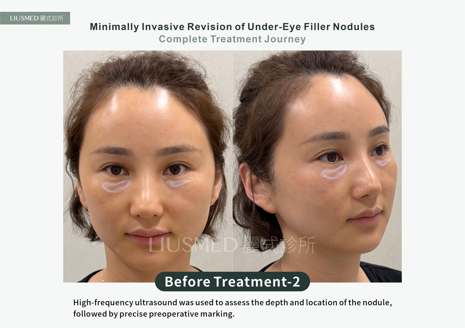
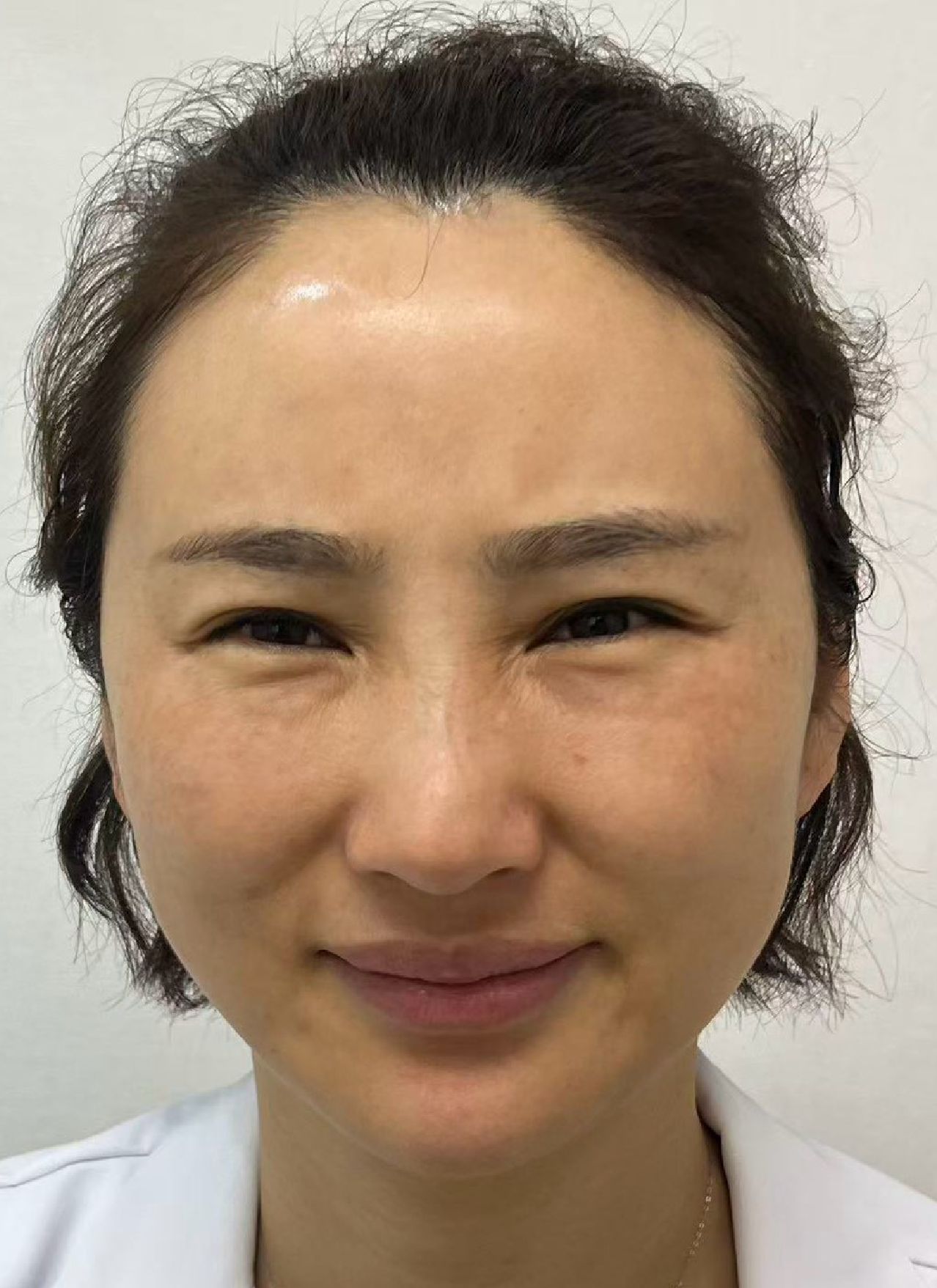
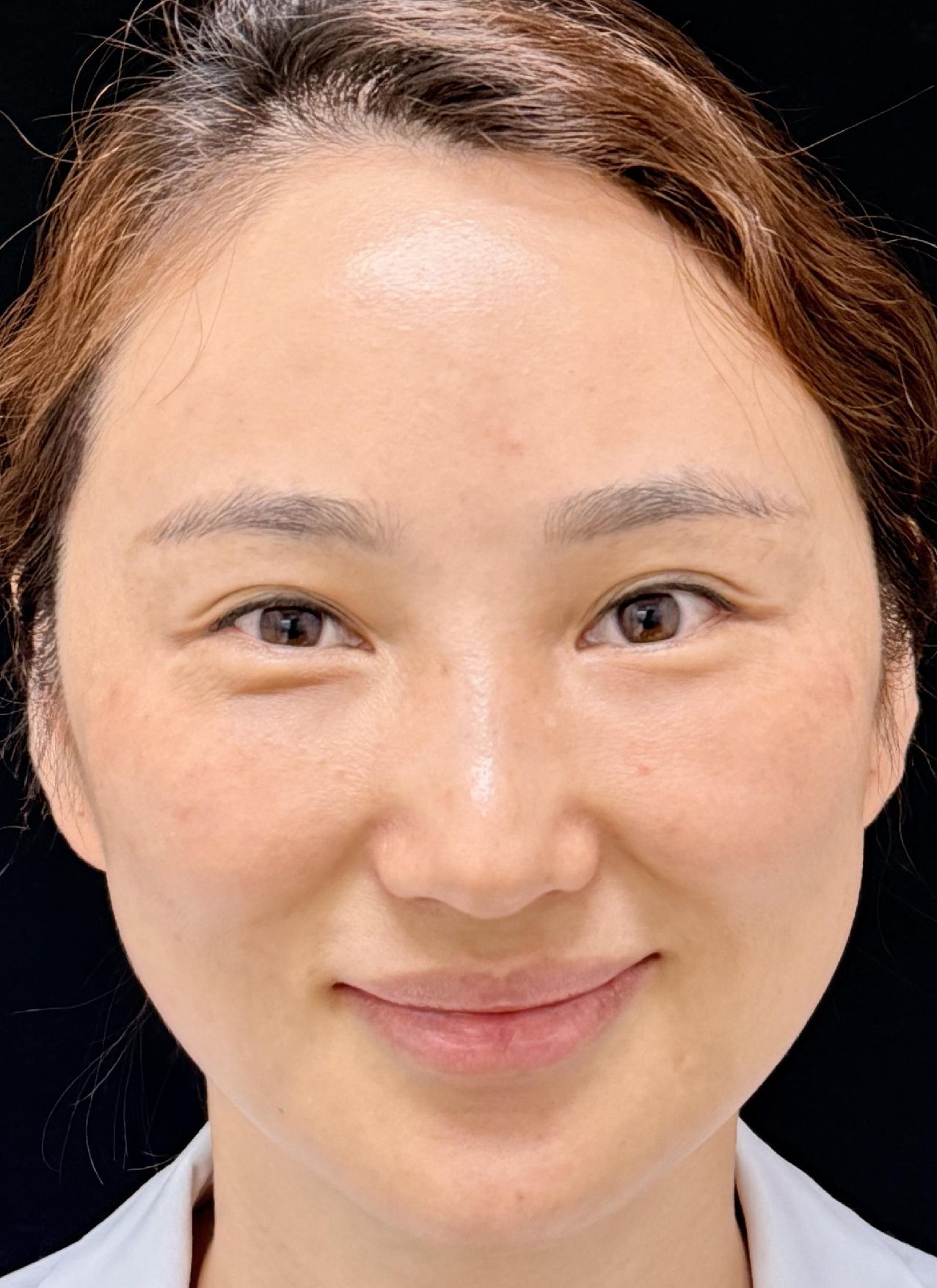
She had autologous fat injected under her eyes to fill a tear trough. The fat did not settle evenly — it consolidated into a lump that felt firm and threw a visible bulge under raking light. To "cover it flat," a collagen stimulator was later injected into the hollow beside it. The result was not a flat under-eye; it was a second lump. Both sat there for more than five years: visible at rest, and pushed into sharper relief whenever the orbicularis muscle contracted in a smile. The question she heard most often from friends was simply, "What happened under your eyes? What did you have done?" The real fork in this patient's road was not the day of removal — it was the moment before the second injection. Once there is already a bulge under the eye, filling the hollow next to it does not level anything; it just adds one more thing that has to be dealt with. And neither autologous fat nor a collagen stimulator has a dissolving enzyme, so there is no way back out of what was injected. This is the most common detour in under-eye work: treating "add more" as the only tool on hand. The first step was not a needle but a look. High-frequency ultrasound mapped the depth, extent and layer relationship of the two masses — which one sat superficially, which lay deeper, where the border between them ran — and the approach was then drawn on the skin before anything was done. The two layers were extracted separately through a pinhole of about 2 mm. At the three-month review the under-eye contour was smooth again, the smile no longer squeezed out two ridges, and scarring was minimal. To be honest about the limits: long-standing material can adhere to surrounding tissue, so complete clearance varies with material, location and time (the first session usually removes about 30–50%, and with most fillers two to three sessions can reach about 80–90%; not a guaranteed 100%), and recovery varies from person to person. (Shared with the patient's consent; images de-identified.)