Clinical Evidence
Our evidence-based approach to filler revision — methodology, outcomes, and the research behind ultrasound-guided extraction.
Our Methodology
Liusmed Clinic employs ultrasound-guided single-pinhole filler extraction, a technique continuously refined by Dr. Ta-Ju Liu over more than 15 years of clinical practice.
- Real-time ultrasound guidance: High-frequency ultrasound precisely locates filler material — its position, extent, and depth — eliminating blind manipulation.
- Single-pinhole approach: Extraction is completed through a single minimal pinhole entry point, resulting in minimal wound, short downtime, and no visible scarring.
- Intraoperative verification: Ultrasound confirmation is performed during and immediately after the procedure to ensure adequate filler removal.
- Multi-material capability: Applicable to hyaluronic acid, Ellanse, Sculptra, Radiesse, silicone, autologous fat, and other filler materials.
Clinical Outcomes
Success definition: "Success" is defined as complete or >90% removal of target filler material, confirmed by post-procedure ultrasound at 1-month follow-up.
Complications: Complication rate <2%, all minor (bruising, temporary swelling). No serious adverse events.
Note: Individual results vary. Statistics reflect aggregate outcomes across all filler types and complication categories.
Evidence Base
The following peer-reviewed publications support the principles underlying our ultrasound-guided filler extraction approach:
Schelke LW, Van Den Elzen HJ, Erkamp PPM, Neumann HAM. Use of ultrasound to provide overall information on facial fillers and surrounding tissue. Dermatol Surg. 2010;36 Suppl 3:1843-1851.
Establishes ultrasound as a reliable imaging modality for detecting and characterizing dermal fillers in facial tissue.
Lemperle G, Gauthier-Hazan N, Wolters M, Eisemann-Klein M, Zimmermann U, Duffy DM. Foreign body granulomas after all injectable dermal fillers: part 1. Possible causes. Plast Reconstr Surg. 2009;123(6):1842-1863.
Comprehensive review of granulomatous reactions to injectable fillers, informing complication classification and management strategies.
DeLorenzi C. New high dose pulsed hyaluronidase protocol for hyaluronic acid filler vascular adverse events. Aesthet Surg J. 2017;37(7):814-825.
Landmark protocol for managing vascular occlusion from HA fillers, establishing the urgency framework for filler emergencies.
Beleznay K, Carruthers JDA, Carruthers A, Mummert ME, Humphrey S. Delayed-onset nodules secondary to a smooth cohesive 20 mg/mL hyaluronic acid filler: cause and management. Dermatol Surg. 2015;41(8):929-939.
Documents delayed filler complications and the longevity of HA fillers beyond expected duration, supporting the need for imaging-guided removal.
Lim TS, Wanitphakdeedecha R, Suphatsathienkul P, Eimpunth S, Manuskiatti W. Facial overfilled syndrome (FOS): a review of the literature on clinical features and grading scale. J Cosmet Dermatol. 2023;22(9):2380-2388.
Defines and categorizes Facial Overfilled Syndrome (FOS), providing a standardized grading system for filler overfilling severity.
Statistical Methods
The ">95% success rate" cited on this page is calculated based on the following methodology:
- Sample size: Consecutive cases of ultrasound-guided filler extraction performed at Liusmed Clinic from 2010 to 2025, totaling over 3,000 cases.
- Follow-up period: Ultrasound follow-up examination at 1 month post-procedure, with select cases followed up to 6 months.
- Success measurement criteria: Complete or >90% removal of target filler material confirmed on post-procedure ultrasound, verified by both the treating physician and an independent ultrasound examiner.
- Exclusion criteria: Cases where treatment was discontinued due to patient factors or where follow-up was not completed are excluded from the statistics.
Conflict of Interest Disclosure
The clinical outcome data presented on this page is derived from Liusmed Clinic's own practice records. This data has not been independently audited by a third party or published in a peer-reviewed journal. Liusmed Clinic provides the filler revision treatments described herein and has a financial interest in these services. Patients should consider this context when evaluating the information presented. We encourage prospective patients to seek independent medical opinions and to review the peer-reviewed literature cited above.
Featured Poster
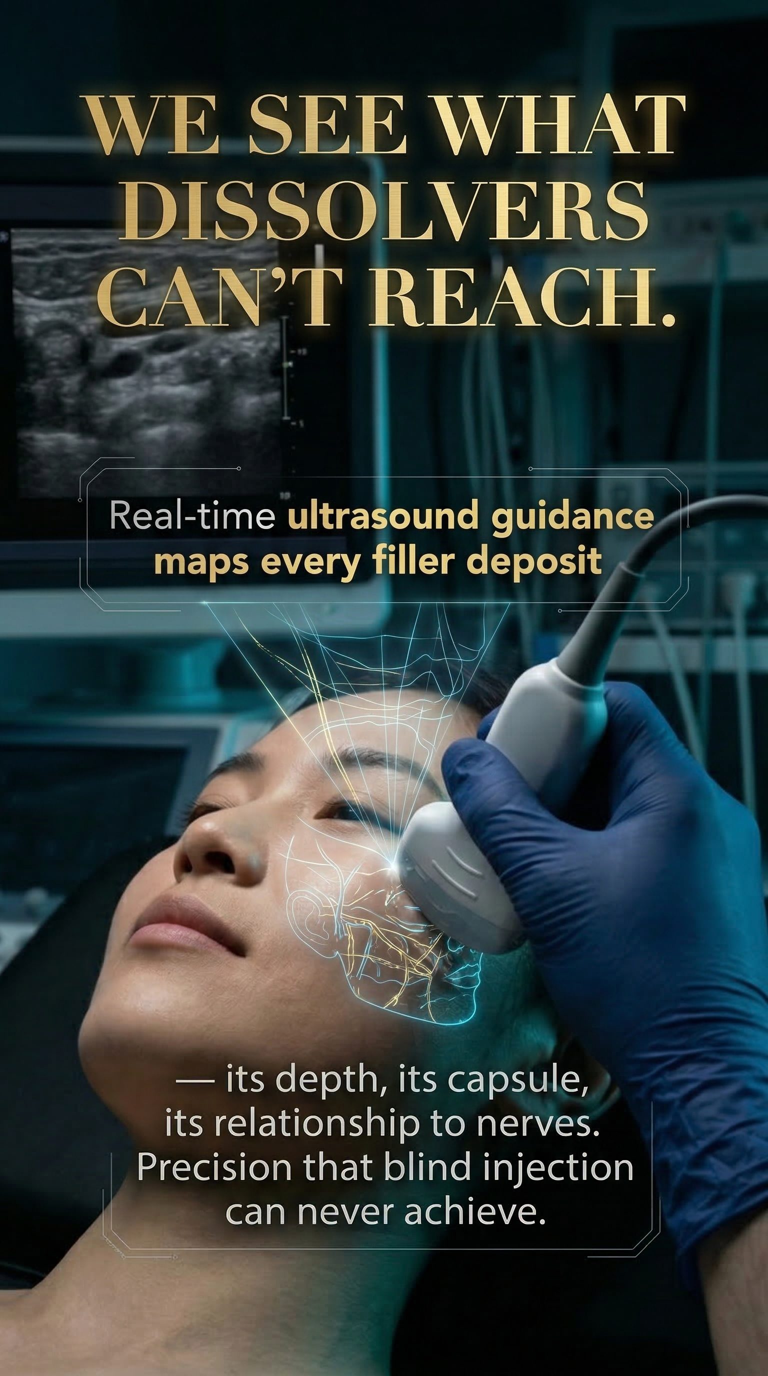
We see what dissolvers can't reach.
Real-time ultrasound guidance maps every filler deposit — its depth, its capsule, its relationship to nerves.
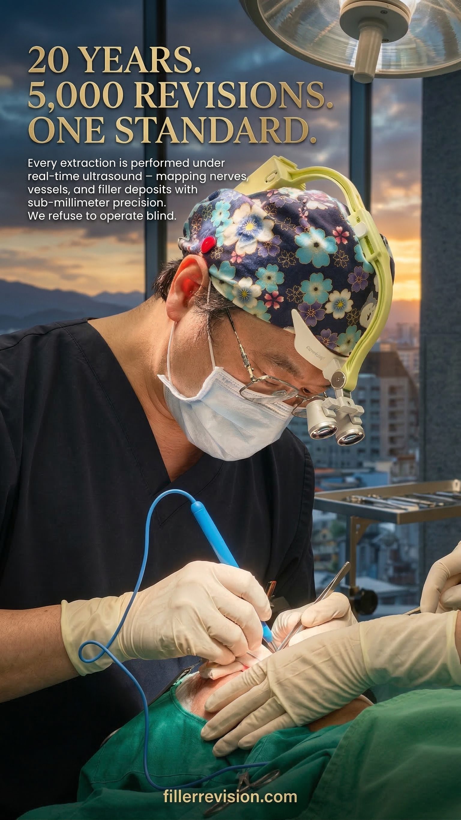
20 years. 5,000 revisions. One standard.
Every extraction is performed under real-time ultrasound — mapping nerves, vessels, and filler deposits with sub-millimeter precision.