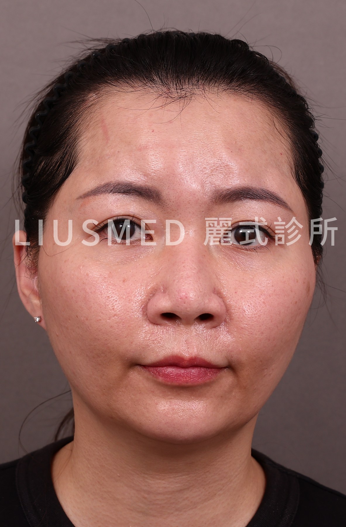
"The news said hyaluronic acid, but I think what I had was AestheFill. Does that make a difference?"
It does, and the difference is large.
That September report about an aesthetic-industry manager who lost function in her left eye after an injection circulated almost entirely under the words "hyaluronic acid," while the article itself described AestheFill. In Taiwan, nicknames push several completely different materials under the popular umbrella term for hyaluronic acid. Day to day that is only messy naming. Once the material is inside a vessel, though, its name stops being a small matter, because HA injected into a vessel has something available to reach for. Biostimulators do not.
The headline says HA; the syringe often held something else
Mapping the nicknames back to their compositions is the foundation for everything that follows.
- AestheFill is the subdermal filler whose main component is PDLLA (poly-D,L-lactic acid), made by Regen in Korea.
- Ellansé, known locally as the "girl needle," is PCL (polycaprolactone).
- Sculptra, the "baby-face needle," is PLLA (poly-L-lactic acid).
- Radiesse, sold in Taiwan as a crystal-clear volumiser, is CaHA (calcium hydroxylapatite).
- Hyaluronic acid (HA) is the only one of these that is actually hyaluronic acid.
Those five lines read like rote memorisation, but the clinic really does get stuck here. Someone arrives with a receipt that says "baby-face needle" and I have to work out whether that meant Sculptra or Juvelook. Someone else only remembers "the one that grows collagen." When the material cannot be identified, management becomes guesswork, which is why I put ultrasound material identification ahead of any procedure.
So why does composition matter so much specifically in vascular occlusion?
Hyaluronidase recognises HA. It does not recognise PDLLA, PLLA, or CaHA
Hyaluronidase is a narrow tool: it cleaves the sugar chains of hyaluronic acid. Once an HA filler is cut apart it becomes small molecules the body clears, which is why it helps both inside and outside the vessel. That is how it became the standard response to HA occlusion.
PDLLA, PLLA, and PCL are solid polyester microparticles. CaHA is a calcium-containing mineral microsphere. Hyaluronidase does nothing to their structure. This is not a dosing problem and not a technique problem, the enzyme simply does not recognise the material.
This is not my personal reading. A 2024 multicentre report in the International Journal of Dermatology opens its abstract with exactly this point: hyaluronidase can dissolve hyaluronic acid-based fillers during vascular adverse events, while non-HA fillers lack such an "antidote" (Kadouch J, et al. Int J Dermatol. 2024;63(9):1252-1255. PMID: 38955457).
← Swipe to see more →
| Material (common nickname) | Composition | Does hyaluronidase dissolve it? | What the literature does during occlusion |
|---|---|---|---|
| Hyaluronic acid | HA | Yes | High-dose, repeated, image-guided hyaluronidase |
| AestheFill ("elf needle") | PDLLA | No | No dissolution route; treat blood flow and inflammation |
| Ellansé ("girl needle") | PCL | No | As above |
| Sculptra ("baby-face needle") | PLLA | No | As above |
| Radiesse (CaHA volumiser) | CaHA | No | As above |
| HArmonyCa | HA + CaHA | Only the HA half | Semi-reversible; the CaHA stays where it is |
Key point: Reversibility is a slowly irritating question when the problem is a lump. When the problem is vascular occlusion, it is the question that decides what can be done that day. Under the same time pressure, HA has a route to take and the other materials require an entirely different line of thinking.
I break down the reversibility differences on the lump side in more detail in 7 reasons hyaluronidase fails. This article stays with the acute phase.
No antidote does not mean nothing can be done
This is the part I most want to be clear about, because "no antidote" is easily read as "nothing can be done," and that is not what the literature shows.
The Kadouch report collected three vascular adverse events caused by non-HA fillers: two from CaHA and one from PLLA. The treatment was ultrasound-guided hyaluronidase injection, and all three resolved without necrosis and without scarring. The explanation the authors offer is not that hyaluronidase dissolved the material, but that vasoconstriction is the primary mechanism: injection-induced spasm holds the perforator arteries constricted for a prolonged period, and relaxing those vessels resolves the picture (PMID: 38955457, DOI: 10.1111/ijd.17355).
On the CaHA side, a 2022 systematic review gathered the CaHA vascular complications in the literature into 43 cases. Eight of them received hyaluronidase, and the reviewers write directly that there is no evidence hyaluronidase can break down or dissolve CaHA fillers. They read the practice as injectors borrowing the drug's oedema-reducing and anti-inflammatory properties rather than any ability to dissolve calcium. Interestingly, the same review still recommends 600 units of hyaluronidase per 0.1 mL of CaHA immediately after a skin ischaemic event (Plast Aesthet Res. 2022. DOI: 10.20517/2347-9264.2022.09).
PCL is on record too. A 2025 case report dealt with vascular occlusion after PCL filler was injected into the nasolabial folds, and the authors used a full combination: hyaluronidase, a steroid (triamcinolone acetonide), low-molecular-weight heparin, antibiotics, and platelet-rich plasma, followed later by fractional CO2 laser for the residual pigment and scarring. At five months the result was minimal scarring and a good cosmetic outcome (PMID: 39757792, DOI: 10.1111/jocd.16779).
All three sources sit at the level of case reports and small series, and the literature is still accumulating a standardised protocol, so I will not present them as a settled pathway. They do point the same way, though: in non-HA filler occlusion, what is being treated is the response of the vessels and tissue, not the material. And all of them assume the same precondition, that the work is done under imaging.
That is why, on the vascular occlusion repair side of the practice, I treat ultrasound as the starting point rather than an optional extra. Without seeing the course of the vessels and the layer the material sits in, you are left extrapolating from anatomical averages, and the difficult thing about the facial danger zones is precisely that vascular courses differ from person to person.
Key point: In non-HA filler occlusion the priority is restoring blood flow to that territory and controlling the inflammatory response, not finding a way to break the material down. Aim at the wrong target and the time is gone.
Signs that mean stop the needle now
← Swipe to see more →
| Sign during or just after injection | What it may mean | What to do |
|---|---|---|
| Pain out of proportion to the volume injected | Raised intravascular pressure or ischaemia | Stop the injection and tell the injector |
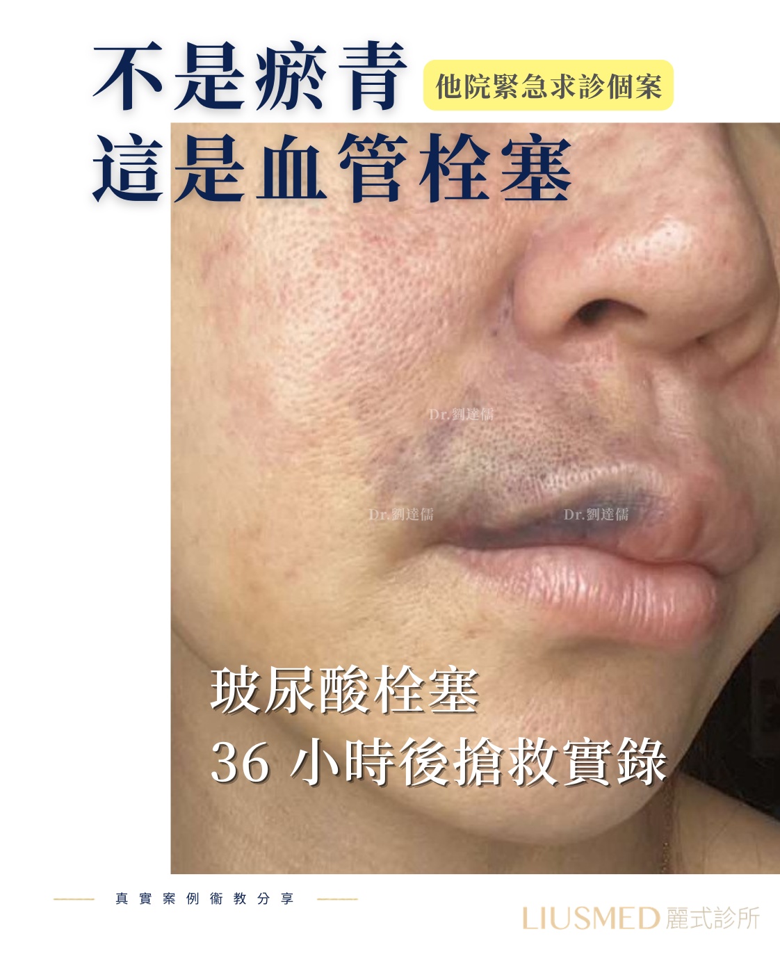
| Blanching, later turning to a mottled purple pattern | Blood flow to that territory is obstructed | Seek care immediately; do not go home and wait |
| Blurred vision, a missing patch of visual field, loss of vision in one eye | Ocular blood flow is affected | Seek care immediately and ask for ophthalmology |
| Pain on moving the eye, a drooping upper lid | Periocular blood flow or nerve involvement | Seek care immediately |
| Colour change or crusting appearing days later | Tissue has moved into post-ischaemic change | Still go in, to assess the extent of injury |
Time is the expensive part here. Occlusion is not an event measured in weeks or even days; it runs in hours, and anything involving the eye is more urgent still. I know the first instinct for many people is to go home and ice it, but this is one of the few situations where I will tell you directly not to wait. The full timeline and emergency principles are in the complete guide to filler vascular occlusion.
The forehead, nose, and tear trough get flagged repeatedly because their vascular networks connect with the supply to the eye. That does not mean injecting there will go wrong; it means the consequences of it going wrong are different.
Who is holding the needle decides whether anyone can help in that moment
Injecting filler is a medical act, and under Taiwan's Physicians Act only a physician may perform it. An injection carried out in a studio, a private home, or a hotel room already fails that standard legally. The more practical problem is that occlusion has to be recognised on the spot.
In those first tens of minutes, the result turns on a short list: whether anyone present understands what blanching and disproportionate pain mean, whether there is an ultrasound to confirm flow, whether the drugs are on hand, and whether there is an ophthalmology or emergency route to transfer into immediately. None of that appears automatically because the person injecting has a steady hand.
I want to say one thing honestly: physicians in legal practice also encounter occlusion. It is a known risk of injectable treatment, not something that happens only with unlicensed injectors. The difference lies in preparedness and in how quickly it is handled afterwards. So I suggest getting a few questions answered before the injection; I have collected them in how to choose a revision physician, and that same list works just as well for deciding whether to let a given person inject you at all.
For those who have already had the injection and are now worried: if you are not even certain what the material was, the first step is not deciding whether to remove it, but establishing what is in there and in which layer. Collagen biostimulator mechanisms and risks fills in some background. If there is already a lump, a distortion, or a colour change, the assessment pathway on the filler revision side starts with imaging rather than with a treatment menu.
To have Dr. Ta-Ju Liu's team assess your case, go to book a consultation.
Frequently Asked Questions
I had AestheFill and there is a pale patch of skin now, but it does not really hurt. Should I go in immediately?
Yes. Painless does not mean fine; blanching means blood flow to that territory is already affected. AestheFill is PDLLA, so there is no enzyme that dissolves it, and everything that can be done is tied to time. The earlier it is addressed, the more options there are.
If biostimulators have no antidote, is hyaluronidase completely useless for them?
Not completely useless, but the reason differs from what you may expect. It will not dissolve PDLLA or CaHA. The effect observed in the literature looks more like relaxing spasmed vessels and reducing swelling and inflammation. That evidence sits at the case-report and small-series level, so whether to use it and at what dose is a physician's judgment under imaging, not something to request by the vial at a clinic.
I only noticed the colour was wrong several days later. Is it still worth going in?
Yes. The acute rescue window has passed, but post-ischaemic tissue keeps changing, and infection, crusting, pigmentation, and scarring all still have room to be managed. The work at that point is assessing the extent of the injury and deciding whether the material inside should be addressed.
I have no idea what I was injected with. Can it still be managed?
It can, and this is a very common situation in clinic. Different materials have different appearances on ultrasound; the echo patterns of hyaluronic acid, polyester microparticles, and calcium microspheres can be told apart. Characterise first, then discuss management. That order cannot be reversed.
References
- Kadouch J, Schelke L, Groh O, Sokol V, Velthuis P. Intralesional hyaluronidase injection to relieve non-hyaluronic acid filler-induced vascular adverse events. Int J Dermatol. 2024;63(9):1252-1255. PMID: 38955457. DOI: 10.1111/ijd.17355.
- Management of vascular complications following calcium hydroxylapatite filler injections: a systemic review of cases and experimental studies. Plast Aesthet Res. 2022. DOI: 10.20517/2347-9264.2022.09.
- Case Report: Management of Polycaprolactone-Based Dermal Filler-Induced Vascular Occlusion. J Cosmet Dermatol. 2025. PMID: 39757792. DOI: 10.1111/jocd.16779.
Content review statement: This article was written by Dr. Ta-Ju Liu based on clinical experience and the existing literature, for health-education reference; it cannot replace an in-person consultation. The management approaches cited here for non-HA filler occlusion come largely from case reports and small series, the evidence is still accumulating, and they do not represent a settled standard protocol. Whether any enzyme, medication, or surgical measure is used still requires case-by-case judgment following an in-person consultation and imaging assessment. If you suspect vascular occlusion after an injection, seek medical care immediately.