The Emergency That Should Never Happen but Must Be Prepared For
If you have experienced sudden skin blanching, severe pain, or color changes during or after a filler injection, you may have encountered — or narrowly avoided — vascular occlusion. This is the most serious complication in filler medicine, and understanding the mechanism is essential whether you are dealing with its aftermath or want to understand the risks before future treatment.
Understanding this process is not meant to create panic, but to serve a practical purpose: the earlier you recognize signs of occlusion, the better the chance of saving tissue.
Key Insight: At FILLER REVISION, our clinical experience confirms that vascular occlusion rescue is a race against time. From skin blanching (ischemia sign) to tissue necrosis (irreversible damage), the window may be only 4-6 hours — even shorter in some areas. This is why we emphasize prevention through ultrasound-guided injection and rapid-response protocols for emergency situations.
Warning Signs to Recognize Immediately
If you have just received a filler injection or are within hours of treatment, the following five signs may indicate vascular occlusion (Vascular Occlusion) and require immediate medical attention — do not wait to see if symptoms resolve on their own:
- Disproportionate, intense pain that feels far worse than normal injection soreness — described by patients as "burning," "electric shock," or "deep aching"
- Sudden skin blanching — a white, pale patch appearing in or near the injection area, sometimes spreading outward in minutes
- Skin color changes — blanching followed by mottled blue, purple, or gray discoloration (Livedo Reticularis pattern is a key warning sign)
- Vision changes — blurred vision, double vision, partial or complete vision loss, eyelid drooping, or eye pain (any vision symptom after facial filler is a true emergency)
- Numbness, weakness, or facial drooping — these may indicate filler has reached arteries supplying nerves or even the brain
What to do in the next 5 minutes: Contact the injector immediately. If you cannot reach them within 10 minutes, go directly to an emergency room and tell them you suspect filler vascular occlusion. Bring the filler brand name and injection site information if possible. Time is tissue — every 30 minutes of delay reduces the salvage rate dramatically (see the timeline table below).
Key Insight: Patients who survive vascular occlusion with full tissue recovery almost always sought help within the first hour. Those who waited "to see if it would get better" frequently developed permanent scarring or tissue loss. When in doubt, treat as an emergency.
Pathophysiology of Vascular Occlusion
Two Occlusion Mechanisms
Direct intravascular embolism: The needle tip penetrates the arterial wall, and filler enters the arterial lumen under injection pressure, physically blocking blood flow downstream. This is a recognized mechanism of vascular compromise from soft-tissue fillers, documented in a clinical case series on filler-related vascular compromise (Beleznay et al., 2014).
Extravascular compression: Large filler volumes compress vessels externally. When external pressure exceeds intravascular pressure, blood flow is blocked.
← Swipe to see more →
| Comparison | Direct Arterial Embolism | Extravascular Compression |
|---|---|---|
| Mechanism | Filler enters vessel lumen | Filler compresses vessel externally |
| Onset speed | Immediate or within seconds | Minutes to hours |
| Ischemia distribution | Along arterial territory | Local compression area |
| Severity | Usually more severe | Depends on compression degree |
| Treatment focus | Dissolve intravascular filler | Decompress (remove/disperse filler) |
| Blindness risk | Present (if ophthalmic artery affected) | Very low |
From Blanching to Necrosis: The Clinical Timeline
Phase 1: Blanching (0-30 Minutes)
Blood flow is blocked; downstream tissue suddenly loses oxygen supply. Skin turns pale, with severe disproportionate pain as the key warning sign.
What can be done: Stop injection immediately. For HA (Hyaluronic Acid) fillers, inject large volumes of hyaluronidase following a high-dose pulsed protocol as described by DeLorenzi, 2017. Apply warm compresses. Administer oral aspirin. Apply nitroglycerin paste topically.
Phase 2: Ischemic Progression (30 Minutes-6 Hours)
Tissue begins anaerobic metabolism. Lactate accumulates. Cell membranes fail. Blanching areas gradually turn dark purple or blue-gray. Pain intensifies. Blisters may appear.
Phase 3: Tissue Necrosis (6-24 Hours)
Beyond the critical ischemia threshold, cells undergo irreversible death. Skin turns deep purple or black. Eschar forms.
Phase 4: Demarcation and Repair (Days-Weeks)
Necrotic tissue separates from viable tissue. Debridement, wound care, and reconstruction may be needed.
Emergency Rescue Principles
Time Is Tissue
← Swipe to see more →
| Time Window | Tissue Status | Salvage Chance |
|---|---|---|
| 0-30 minutes | Ischemic but reversible | High (>80%) |
| 30 min-2 hours | Worsening ischemia | Moderate (50-80%) |
| 2-6 hours | Partial cell death beginning | Limited (20-50%) |
| 6-12 hours | Extensive necrosis progressing | Very low (<20%) |
| >12 hours | Irreversible necrosis | Damage control only |
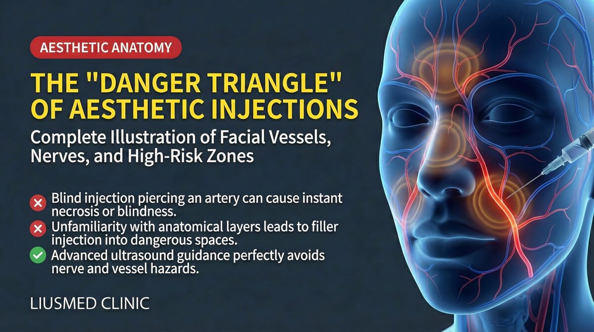
Key Insight: Blindness is the most irreversible outcome. Once filler reaches the central retinal artery, even immediate treatment has very low probability of restoring vision. Prevention — through ultrasound guidance, avoiding danger zones, slow small-volume injection — is the most important strategy. Learn about facial anatomy danger zones.
Clinical Implications for Revision Patients
For patients who have survived a vascular occlusion event, the aftermath often involves tissue damage that requires careful management — and an understanding of what caused the event in the first place. At FILLER REVISION, we see patients dealing with post-occlusion scarring, residual filler that contributed to the vascular compromise, and the anxiety of knowing filler remains near critical vessels. Ultrasound (Ultrasonography)-guided assessment can identify remaining filler deposits in relation to the vascular anatomy, helping determine whether residual material poses ongoing risk and whether removal is advisable. For patients considering future injectable treatments after a vascular event, this imaging-based understanding of their anatomy is essential for making safe decisions.
Special Area Risks
Nasal Occlusion
The nose is one of the most common sites for filler-related vascular occlusion. Patients with nose filler displacement may have experienced mild vascular compression events without realizing it.
Ophthalmic Artery Occlusion and Blindness
The most catastrophic outcome: retrograde embolization to the ophthalmic artery. The retina is extremely ischemia-sensitive — central retinal artery occlusion can cause permanent vision loss within 60-90 minutes.
Real case → 36-hour delayed vascular rescue via ultrasound-guided IAHA — a documented success at the rescue window most clinics consider unsalvageable.
The Importance of Prevention
Occlusion rescue is never as effective as prevention:
- Ultrasound-guided injection: Identify vessel locations in real time
- Understanding facial anatomy danger zones
- Small-volume, slow injection: Reduce injection pressure to lower retrograde embolization risk
- Aspiration test: Aspirate before injection to check needle tip is not intravascular
- Cannula use: When appropriate, to reduce vessel puncture risk
- Emergency drugs ready: Hyaluronidase, nitroglycerin always available
Learn about the filler repair evaluation process and our vascular occlusion treatment services. At FILLER REVISION, we believe the best vascular occlusion outcome is the one that never happens — and when it does, rapid recognition and evidence-based intervention save tissue and lives.
Frequently Asked Questions
How soon after a filler injection does vascular occlusion start — can it be delayed?
Most vascular occlusions announce themselves immediately or within minutes of injection — sudden, disproportionate pain and skin blanching are the classic early signs. Occlusion can also evolve over several hours, particularly with extravascular compression, where gradual swelling closes off a vessel. Delayed presentations a day or more later are uncommon for true arterial embolism but can occur with progressive compression or biofilm-related swelling. The practical rule: any disproportionate pain, blanching, or dusky discoloration in the hours after filler should be treated as vascular compromise until proven otherwise — do not wait for it to "declare itself."
How do I tell the difference between vascular occlusion and normal post-injection swelling?
Normal post-injection swelling is symmetric, mild, painless or only slightly tender, and appears within minutes evenly across the treated area. Vascular occlusion is asymmetric, intensely painful out of proportion to what was done, and involves color change — blanching, dusky purple, or grayish patches that follow a streaky pattern along an artery's course. The most reliable distinguishing feature is pain that worsens rather than settling, combined with any color change. If you are uncertain, photograph the area every 15 minutes and compare — true occlusion changes visibly within an hour. When in doubt, contact your injector or go to the emergency room.
I just noticed pale or dusky patches several hours after my injection — is it too late to do anything?
It is not too late, but every hour matters. Tissue salvage rates drop from over 80% in the first 30 minutes to roughly 20–50% by 6 hours, and below 20% after 12 hours of ischemia. Even at 12–24 hours, prompt hyaluronidase (Hyaluronidase) administration (for HA fillers) and supportive treatment can still limit the necrotic zone and reduce permanent scarring. Go to an emergency room or contact your injector immediately — never assume "it's been too long, nothing will help." Emergency rooms with experience in cosmetic complications can also coordinate with plastic surgery for tissue reconstruction if necrosis has begun.
After surviving a vascular occlusion, can I ever safely have filler again?
Many patients do — but only with significantly increased safety precautions and ideally not in the same anatomical area. The decision depends on three factors: (1) how the occlusion happened (technique error, anatomical variation, or unavoidable bad luck), (2) what filler and area you are considering, and (3) whether the injector uses ultrasound (Ultrasonography) guidance. After a vascular event, ultrasound-guided injection is no longer optional — it is essential. At FILLER REVISION, we evaluate post-occlusion patients with ultrasound to map their actual vascular anatomy, identify residual filler that may still pose risk, and discuss whether future injectables can be done safely. For some patients, the safer answer is to switch to non-injectable alternatives entirely.
Key Insight: Vascular occlusion is a war against time, but the best battle is the one that never begins. "Seeing" vessels under ultrasound before injecting is far better than starting rescue after occlusion occurs. Prevention is always more effective than treatment.