Every Needle Passes Through a Complex Anatomical Maze
If you are dealing with a filler complication — or trying to understand why one occurred — it helps to know what lies beneath the surface where your filler was placed. You may have been told the injection was routine, but beneath every injection site lies an extraordinarily complex three-dimensional anatomical structure: arteries coursing through multiple layers, veins forming dense networks, and nerve branches distributed like wiring throughout every tissue plane.
With each injection, the needle tip navigates blindly through this structure — unless you can "see" everything beneath the skin.
Key Insight: At FILLER REVISION, our clinical experience confirms that facial vascular anatomy shows significant individual variation. The "standard anatomy" in textbooks represents only statistical averages — your blood vessels may course in entirely different positions. This is why even experienced injectors can encounter unexpected complications, and why ultrasound-guided assessment is essential for both injection and revision.
The Basic Architecture of the Facial Vascular System
Major Arterial Sources
The facial blood supply comes primarily from two arterial systems:
External carotid artery system:
- Facial artery: Ascends along the mandibular border, passing the oral commissure and nasal ala, reaching the medial canthus (becoming the angular artery)
- Superior and inferior labial arteries: Branch from the facial artery, forming circular anastomoses in the lips
- Superficial temporal artery: Supplies the temple and lateral forehead
Internal carotid artery system:
- Ophthalmic artery: Forms extensive anastomotic networks with facial vessels through intraorbital branches
- Supratrochlear and supraorbital arteries: Supply the central and upper forehead
The Dangerous Anastomotic Network
The most critical anatomical fact: extensive anastomoses exist between the external and internal carotid artery systems. Cadaveric studies have mapped these facial danger zones in detail, identifying the specific arterial pathways through which filler can travel (Tansatit et al., 2015). This means filler that accidentally enters a facial artery can theoretically travel retrograde through these networks to reach the ophthalmic artery, causing vascular occlusion or even blindness.
← Swipe to see more →
| Danger Zone | Vessels Involved | Primary Risk | Risk Level |
|---|---|---|---|
| Glabella | Supratrochlear, supraorbital arteries | Skin necrosis, blindness | Very high |
| Nose | Dorsal nasal, lateral nasal arteries | Skin necrosis, blindness | Very high |
| Nasolabial fold | Facial artery, angular artery | Skin necrosis | High |
| Temple | Superficial temporal artery branches | Skin necrosis, stroke | High |
| Tear trough/infraorbital | Infraorbital artery, angular artery | Skin necrosis, vision impairment | High |
| Forehead | Supratrochlear, supraorbital arteries | Skin necrosis | Medium-high |
| Nasolabial folds | Facial artery | Skin necrosis | Medium |
| Lips | Superior/inferior labial arteries | Tissue necrosis | Medium |
Detailed Analysis of High-Risk Zones
The Nose: One of the Most Dangerous Injection Areas
Nasal filler is the most popular non-surgical rhinoplasty method and simultaneously one of the highest areas for vascular occlusion.
Anatomical risk factors:
- The dorsal nasal artery is a terminal branch of the ophthalmic artery that anastomoses with the angular artery of the facial artery
- Dense lateral nasal artery branches around the nasal ala
- Thin nasal skin with minimal subcutaneous tissue allows fillers to easily compress vessels
- Individual vascular variation is particularly prominent in the nose
Nasal filler displacement and complications are common clinical problems. Learn more about nose filler displacement.
The Glabella: A Highway to the Eyes
The supratrochlear and supraorbital arteries in the glabellar region connect directly to the ophthalmic artery. Filler entering these vessels can travel retrograde extremely rapidly to the central retinal artery, causing irreversible blindness. A systematic review of strategies for avoiding and treating filler-induced blindness has underscored the critical importance of this anatomical knowledge (Beleznay et al., 2015).
The Temple: Hidden Risk Zone
Temple filler improves hollowing, but the superficial temporal artery and deep temporal arteries form complex vascular networks in this area. Deep injection may affect vascular branches leading to the meninges.
The Mechanism of Vascular Occlusion
From Injection to Onset
When filler accidentally enters an artery or compresses an external vessel:
- Mechanical obstruction: Filler material physically blocks blood flow
- Vasospasm: Vessel walls contract violently when stimulated, further reducing blood flow
- Retrograde embolization: Under injection pressure, filler may travel against blood flow direction to reach distant branches
- Secondary thrombosis: After blood flow stasis, clotting factors activate, forming thrombi that expand the blockage area
Key Insight: Vascular occlusion is a medical emergency. From the appearance of skin blanching and pain to tissue necrosis, there may be only hours in the rescue window. Learn more about the golden time for vascular occlusion emergency treatment.
Why Anatomy Knowledge Matters for Revision Patients
For patients presenting at FILLER REVISION with complications, understanding facial anatomy explains not only what went wrong but also why safe revision requires the same anatomical precision — if not more. Correcting displaced or problematic filler means working in tissue that has already been altered by previous injections, inflammation, or scarring. The vascular and nerve structures are still there, and in some cases, their positions may have shifted due to the filler itself. This is why every revision procedure at FILLER REVISION begins with ultrasound mapping of both the filler deposits and the surrounding vascular anatomy. Navigating these danger zones safely during revision is just as important as avoiding them during the original injection.
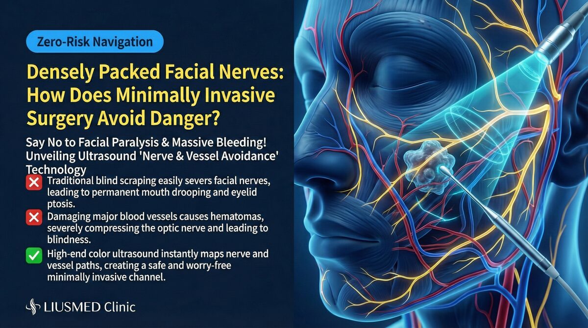
Nerve Distribution and Injection Risks
Beyond vascular risks, facial nerve distribution must also be well understood:
Three Branches of the Trigeminal Nerve
- Ophthalmic nerve (V1): Supplies sensation to forehead and upper eyelid
- Maxillary nerve (V2): Supplies sensation to mid-face (cheek, nasal ala, upper lip)
- Mandibular nerve (V3): Supplies sensation to lower face and motor function to masticatory muscles
Facial Nerve Branches
The facial nerve controls facial expression muscles. Its branches are particularly superficial in the preauricular and buccal regions, making them vulnerable to injection-related injury that may cause temporary or permanent facial asymmetry.
← Swipe to see more →
| Nerve Injury Type | Symptoms | Recovery Time |
|---|---|---|
| Temporary sensory numbness | Decreased sensation at injection site | Usually weeks to months |
| Temporary motor injury | Local expression muscle weakness | Usually weeks |
| Permanent sensory injury | Persistent abnormal sensation | May not fully recover |
| Permanent motor injury | Permanent facial asymmetry | May require surgical repair |
Real case → 36-hour delayed vascular rescue via ultrasound-guided IAHA — nasolabial fold vascular occlusion where ultrasound mapping of facial artery branches enabled rescue at a window most clinics consider unsalvageable.
How Ultrasound (Ultrasonography) Changes Injection Safety
"Seeing" Beneath the Skin
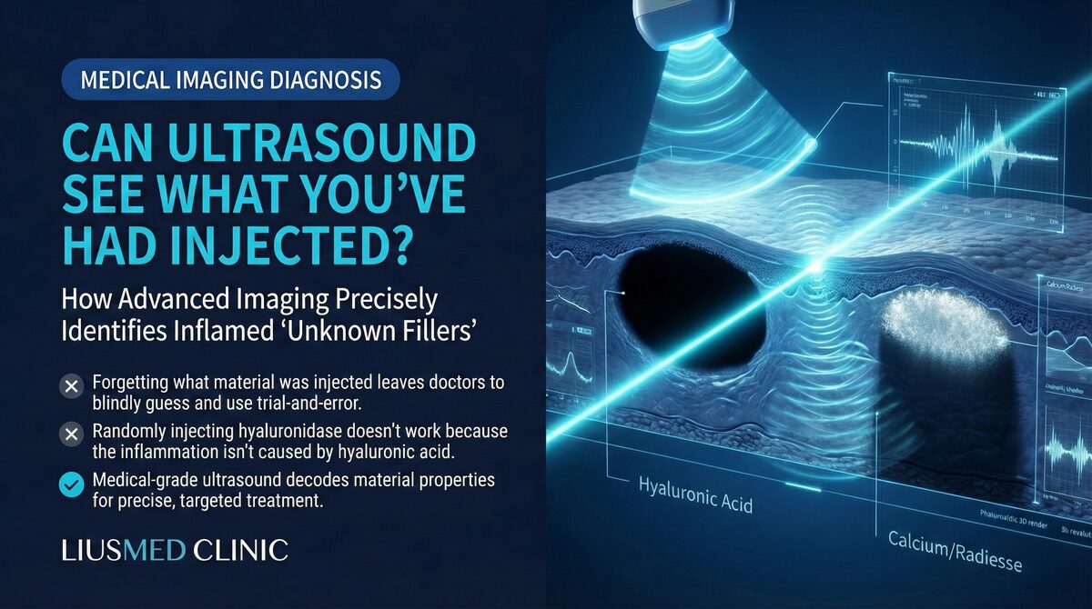
High-resolution ultrasound provides real-time anatomical information before, during, and after injection:
Pre-injection assessment: Personalized vascular mapping, identification of variant arteries, confirmation of prior filler residue locations, and tissue layer evaluation.
Intra-procedural guidance: Real-time needle tip confirmation, avoidance of identified vessels, monitoring of filler spread direction, and early detection of abnormalities.
Post-injection monitoring: Confirmation of final filler distribution, assessment for signs of vascular compression, and baseline data for future adjustments.
← Swipe to see more →
| Comparison | Blind Injection | Ultrasound-Guided |
|---|---|---|
| Vessel identification | Relies on anatomical knowledge | Real-time visualization |
| Individual variation | Cannot confirm | Can identify |
| Filler localization | Tactile judgment | Precise positioning |
| Old residue detection | Impossible | Detectable |
| Early complication detection | Difficult | May detect before symptoms |
Understand Your Risk, Make Informed Choices
Facial anatomical complexity reminds us that filler injection is not a "simple lunchtime procedure." Every needle operates in a three-dimensional space filled with vessels and nerves.
Risk reduction strategies:
- Choose an injector with ultrasound capability for safer decisions
- Understand the risk level of your injection area
- Confirm emergency protocols — does your injector have vascular occlusion emergency plans?
- Know when to seek help — immediate medical attention for blanching, severe pain, or vision changes
Filler migration and complications are closely related to anatomical structures. Learn about how fillers migrate and the complete filler repair evaluation process.
If you are facing filler-related concerns or suspect a vascular complication, FILLER REVISION provides ultrasound-guided assessment that maps your unique anatomy alongside existing filler deposits — because safe revision means seeing exactly what is there before taking any action.
Key Insight: The best injection safety strategy is not memorizing "average positions" from anatomy textbooks, but using ultrasound to see in real time where "this person's" vessels actually are. Personalized safety begins with personalized visualization.
Frequently Asked Questions
Which areas of the face are the highest-risk injection zones, and why?
The glabella (between the eyebrows) and the nose are the highest-risk zones because their arteries connect directly to the ophthalmic artery. If filler accidentally enters these vessels, it can travel retrograde to reach the eye, creating a risk of irreversible blindness. The nose is also high-risk because its skin is thin with little subcutaneous tissue, so fillers can easily compress vessels, and vascular variation there is particularly prominent.
What warning signs after a filler injection mean I should seek help immediately?
Seek immediate medical attention if you notice skin blanching (whitening), severe pain, or any vision changes after injection. These can signal vascular occlusion, which is a medical emergency: from the appearance of blanching and pain to tissue necrosis, there may be only hours in the rescue window. Acting quickly matters because the blockage can become irreversible.
If textbooks describe a 'standard' facial anatomy, why do complications still happen with experienced injectors?
Facial vascular anatomy varies significantly between individuals, and the 'standard anatomy' in textbooks represents only statistical averages — your blood vessels may course in entirely different positions. Because the needle navigates blindly beneath the skin, even experienced injectors can encounter unexpected complications. This is why the article emphasizes assessing where your vessels actually are rather than relying on average positions.
How does ultrasound make injection or revision safer compared with blind injection?
High-resolution ultrasound allows the doctor to 'see' beneath the skin in real time, mapping your personal vascular anatomy before injection or revision and identifying variant arteries and prior filler residues that blind injection cannot detect. During the procedure it confirms needle position and helps avoid identified vessels, and afterward it checks the final filler distribution. The article frames this as replacing guesswork with imaging — seeing exactly what is there before taking any action.
Is correcting or removing old filler safer than the original injection?
Not automatically — revision surgery in previously injected tissue carries the same anatomical risks as the original injection. Working in tissue already altered by prior injections, inflammation, or scarring is demanding because the vascular and nerve structures are still present, and in some cases their positions may have shifted due to the filler itself. This is why each revision at FILLER REVISION begins with ultrasound mapping of both the filler deposits and the surrounding vascular anatomy.