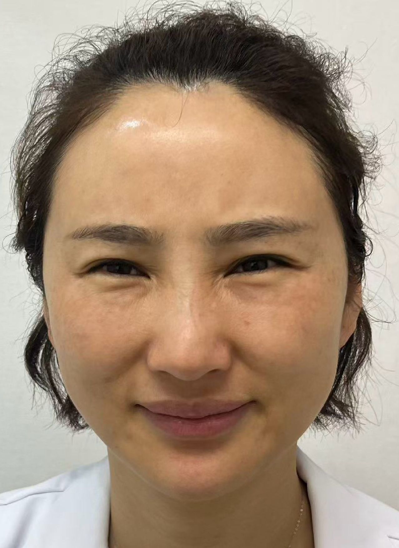
"It has been over a month and it is still dark under here."
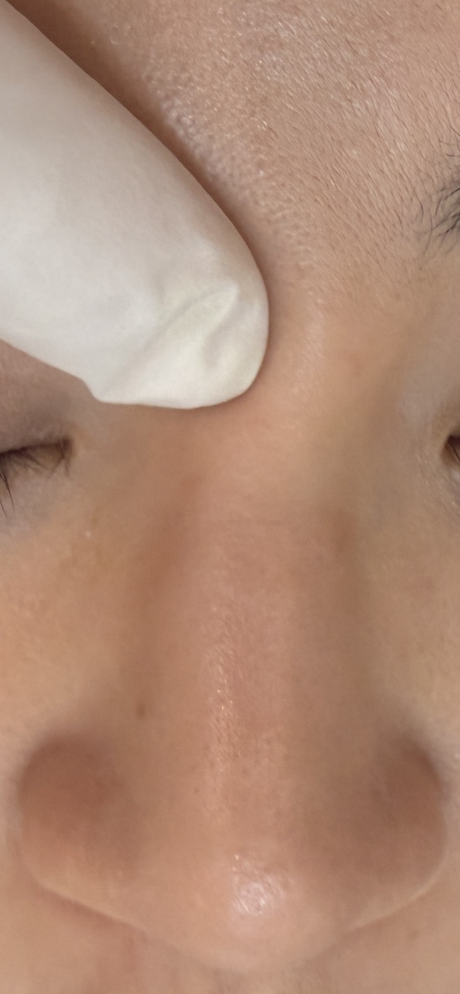
She took her mask off before sitting down and drew a circle under her right eye with a fingertip. It did not look like discrete lumps. It looked like a layer of shade laid over the area, deepest at the side nearest the nose. For the first fortnight she assumed it was a bruise. By the third week it started to bother her, because the colour had stopped changing.
My first question was not whether to treat it. It was what the product was called.
She had to think about it. What she remembered was a nickname, not the registered Chinese name.
That matters more than it sounds. Nicknames appear nowhere in an approved licence. You need the registered name, GOURI, to pull up the licence and read what it actually says. The name people use in the clinic will not find you a single official document.
This article is not about whether you should have had it. You already did. What has to be dealt with is the colour in the mirror, and it could be one of four different things. Get that wrong and every step after it drifts.
First, establish which PCL you were given
Two different products currently travel under the label "liquid PCL" in Taiwan.
GOURI is the genuinely particle-free liquid form. Ellansé is the microsphere form, with PCL (polycaprolactone) spheres suspended in a carrier gel. Same principal polymer, different physical form, different appearance on ultrasound, and different approved sites.
Why can this step not be skipped? Because every reading that follows rests on it. Microspheres give a distinct particulate echo on ultrasound; the liquid form does not, so the reading leans much more on surrounding tissue changes and on capsule behaviour. Even the question "was my injection inside the approved indication" has opposite answers for the two.
How to tell them apart, and how their composition and labelling differ, is set out in "Liquid PCL" means two different products. The material itself is covered on the Ellansé / PCL material page.
What Taiwan approved: lateral canthal lines, 12 weeks, nothing periorbital
GOURI's Taiwanese licence is recent. It was issued on 13 May 2026 under licence number 038200.
The performance section reads, in substance: the product is implanted in the mid to deep dermis to fill facial tissue, for short-term (e.g. 12 weeks) improvement of lateral canthal lines in adults aged 19 and over. In brackets it adds that long-term safety and efficacy remain to be established.
Those few lines settle three separate questions.
← Swipe to see more →
| What the approved labelling says | What it means for you, now that it is in |
|---|---|
| Implanted in the mid to deep dermis | A depth is specified. Placed shallower than that, the optics change |
| Short-term (e.g. 12 weeks) improvement of lateral canthal lines | The approved site is the crow's feet area. Infraorbital, tear trough and the aegyo-sal line are not in it |
| Long-term safety and efficacy remain to be established | For anything measured in months, the approved document itself offers no backing |
One thing needs saying plainly, because it gets read the wrong way. Use outside the approved indication is lawful and common across every speciality, and it does not mean the person who injected you did something wrong. Its meaning is narrower than that: in that region, this product has not completed official safety and efficacy validation. So when something does happen in a region outside the approval, there is less published data available to you than you would expect. This is a question of being informed.
Key point: The approved document will not make the decision for you, but it does mean the sentence "this might be related to that injection" is no longer something you have to argue alone.
Sorting out what the darkness actually is
The mistake I see most often in clinic is treating every kind of under-eye darkness as one thing and waiting it out.
At least four sources are relevant to liquid PCL. They look alike and behave very differently over time.
← Swipe to see more →
| What the darkness is | Where it comes from | Colour and appearance | How it moves over time |
|---|---|---|---|
| Post-injection bruise | Bleeding along the needle path | Purple-red to blue-purple, blurred edges | Changes colour, fades |
| Retained pigment | Breakdown products not yet cleared | Brownish, dull yellow | Fades very slowly, but the direction is lighter |
| Light scattering off the material | Material sitting too shallow, light scattering within it | Blue-grey, a flat muted patch | Does not change on its own while the material stays in that layer |
| Shallow or uneven placement | Position and volume unevenly distributed | Shifts with the light, worst in side light | Moves with the light, not with time |
The last two share a feature: they are not "not yet faded". They are the result of position. If the position does not move, neither does the colour.
There is one more layer. If the skin under your eye already showed vessels or carried pigmented dark circles, filler stretching and thinning the skin will amplify a colour that was always there, and what you see is two things stacked. That layer has nothing to do with which product was used; it belongs to the general differential for under-eye discoloration, which is written up in Still blue after dissolving, including how to separate retained pigment from the Tyndall effect by timing. I will not repeat it here, and will stay with what is specific to liquid PCL.
Key point: A bruise changes colour and fades. Scatter does not. A photograph every two weeks from the same angle in the same light beats anything you can reconstruct from memory.
What the 2026 case series actually recorded
You may reasonably want to know whether anyone has studied this. Someone has, and it is very recent.
In January 2026, Clinical, Cosmetic and Investigational Dermatology published a retrospective case series (PMID 41869420, doi 10.2147/CCID.S571602) on exactly this: bruising that would not clear after liquid PCL injection, in seven cases, all of them GOURI. The authors are spread across two Tokyo clinics, three in Korea, and practices in Southeast Asia, Eastern Europe, China and Malaysia. It is a multi-country collection rather than one country's report.
Here is what it recorded (same paper, PMID 41869420):
← Swipe to see more →
| Item | What the paper records |
|---|---|
| Cases | Seven, retrospectively collected, no control group |
| Sites | Five infraorbital, one at a zygomatic puncture point, one on the upper arm |
| Duration | The abstract says persisting for several months; case by case, the quickest cleared in five days and the two slowest took roughly five to six months from injection to resolution |
| Management | Four mechanisms in parallel: pigment fragmentation with lasers, thermal loosening, mechanical dispersion, and enzymatic degradation |
| Outcome | All patients ultimately achieved resolution, with no serious adverse events |
| Proposed mechanism | Two parallel hypotheses: entrapment of pigment molecules by a dense liquid PCL scaffold, or a Tyndall effect |
Two lines are worth having verbatim. The abstract states that the phenomenon is not permanent, as its resolution parallels scaffold degradation over time. The discussion adds that the Tyndall effect often resembles a mild, deep bruise and is frequently mistaken for one, but that unlike general bruising it remains unchanged over time unless the material is degraded or physically removed.
That second sentence is where the table above comes from, and it is the test you can apply at home.
Now the limits, which you should have as well. Seven cases, retrospective, no control group: this is not high-grade evidence. Three of the thirteen authors are members of the manufacturer's medical research team, and the paper discloses it. The authors also state in their introduction that this is the first report of its kind, so the word "rare" currently has no denominator behind it.
I still cite it, because it is the only document that has recorded this properly. But citing it means carrying those lines along with the findings.
Why "let's try some hyaluronidase and see" is a poor idea here
Hyaluronidase (to be used only after an in-person medical assessment) cleaves the glycosidic bonds of hyaluronic acid. PCL is a polyester, and the enzyme has nothing to grip. That holds for both the microsphere and the liquid form, and it is covered fully in Can hyaluronidase dissolve Ellansé.
So why do people say it worked? Because in that case series one patient did receive hyaluronidase and cleared in six days, the fastest in the group. That case was on the upper arm, not under the eye. The reasonable reading is dispersion of what was retained in the surrounding tissue, or treatment of hyaluronic acid present at the same time — one of the under-eye cases in that series had Restylane injected in the same session. It did not dissolve PCL.
The other line people quote is lipase. The in-vitro data cited in the same paper (discussion section, PMID 41869420) had PCL fully degraded within 120 hours in the presence of lipase. That is a bench result, not a clinical route, and there is a long distance between the two.
Under the eye there is one more layer to this. Hyaluronidase does not distinguish filler hyaluronic acid from the hyaluronic acid in your own tissue. Under-eye skin is measured in fractions of a millimetre and is the thinnest on the face. Whatever extra gets dissolved is written straight onto the face: thinner, more hollow, vessels showing through, and the area looks darker than before.
Key point: Under the eye, a trial injection is not a free experiment. If the direction was wrong, what you lose is thickness you did not have much of.
When to wait, when to have it looked at, when to consider treating
Reasonable to wait: still within the first few weeks, colour still changing, area still shrinking, and it does not stop you going out. Observation here is sound advice rather than a brush-off.
Worth having looked at: a month or two in with no change in colour at all; a blue-grey cast that gets worse in side light; a palpable raised area or firmness; recurring swelling; or the patch becoming more obvious rather than less.
Starting to consider treatment: imaging shows material concentrated in a very superficial layer; two or three approaches at the same site have made no difference; you check the mirror every day to see whether it has grown.
Ultrasound at this stage answers four questions: which layer the material sits in, whether it is diffuse or concentrated, how close it is to the periorbital vessels, and how much skin thickness you still have. The first three decide whether and how anything can be done. The last decides whether you end up more hollow afterwards, and it is the one most often skipped under the eye even though it determines the result.
Honestly, the liquid form is harder to read on ultrasound than the microsphere form. With no spheres there is no clear particulate echo, so the reading depends on surrounding tissue changes and capsule behaviour. I am not going to promise you that it will always be visible. What I will say is that the quality of the decision differs enormously between having looked and not having looked.
When removal is genuinely needed, what we do is ultrasound-guided localisation and extraction of the material and the excess stimulated tissue through a one to two millimetre pinhole. The honest figure is that most cases allow roughly eighty to ninety per cent to be removed, not all of it, and it varies between people. The full approach is on the filler repair service page, and why the under-eye region demands particular restraint is in Under-eye filler extraction.
Before the next injection: what you can actually do
This section is for anyone still deciding, and for anyone who has had it done and wants to lower the odds next time.
To be clear, these lower the odds. They do not guarantee that nothing will happen.
Ask three things: which product this syringe holds, which layer it is going into, and whether this region falls inside its approved indication. The third sounds blunt, but you are entitled to the answer and your doctor has a duty to give it.
Blood-thinning medication and supplements raise the chance of bruising. Whether to adjust anything around a procedure has to be discussed with the doctor who prescribed it. Do not stop medication on your own.
Periorbital skin is thin and densely vascular, so every injection detail is magnified: cannula or needle, the layer entered, the volume at a single point, the speed of injection. Those are the injector's calls, but you can ask about them, and you can cooperate with cold compression and pressure afterwards.
One last thing has nothing to do with technique. If your under-eye area was already dark or your vessels already showed, that base colour will not disappear after an injection and may well be amplified. To work out which type of dark circle you have before anything goes in, the three types of dark circles is a reasonable self-assessment.
Frequently Asked Questions
How long does under-eye bruising after GOURI (liquid PCL) have to last before it counts as abnormal?
Ordinary post-injection bruising fades noticeably and changes colour within two to three weeks. Past a month with no change in colour at all, "still clearing" is no longer a good explanation. The seven-case series from 2026 (PMID 41869420) recorded durations from a few days to roughly five or six months, all of which resolved eventually, but that is a small retrospective series and cannot be used to estimate your own odds.
Mine is blue-grey. Is that still a bruise?
Not necessarily. The Tyndall effect is colour produced by light scattering off the material itself, and it is frequently mistaken for a deeper bruise. The way to separate them is time: a bruise changes colour and fades, while scatter does not change on its own as long as the material stays in that layer. Photographing the area in consistent light is the most practical test.
Would hyaluronidase make it clear faster?
It does nothing to PCL. Hyaluronidase cleaves the glycosidic bonds of hyaluronic acid, and PCL is a polyester, so the chemistry does not match. The case in that series who cleared in six days after hyaluronidase was on the upper arm, and the reasonable reading is dispersion of retained material or treatment of hyaluronic acid injected at the same time, not dissolution of PCL. Under-eye skin is extremely thin, which makes this a poor place for a trial injection.
Will this darkness be permanent?
Based on the only relevant paper to date (PMID 41869420), all seven cases eventually resolved, and the authors consider the phenomenon non-permanent, with resolution tracking the degradation of the material. That is seven cases with no control group, and depth, volume and baseline colour differ between people. The more practical statement is that it usually improves as the material degrades, on a timescale of months.
Can GOURI be injected under the eye in Taiwan? Does having had it mean my doctor was wrong?
The approved Taiwanese indication is short-term (e.g. 12 weeks) improvement of lateral canthal lines, and the infraorbital region and tear trough are not in it. Use outside an approved indication is lawful and common in medicine, and it does not mean your doctor did anything wrong. The practical consequence for you is that no official validation data exists for that region, so both you and your doctor have less evidence to work with if something goes wrong.
Could a laser just remove the colour?
It depends on what is producing the colour. Lasers act on pigment, so retained pigment is a plausible target, while blue-grey from material scatter is a different mechanism altogether. In terms of sequence, working out the source before applying energy is safer than trying one pass to see. Energy around the eye also has to account for distance from the globe and for shielding, which needs an in-person assessment.
There is also a small firm area, not just colour. Is that the same problem?
Not necessarily the same, but worth looking at together. Colour and palpable firmness show up as different findings on ultrasound. When both are present it usually means the material has collected locally, or that surrounding tissue has already reacted. In that situation I would not keep watching the colour alone. Get one imaging assessment first, then decide whether to wait or to act.
Checking where you stand
If you have had GOURI or another liquid PCL and the dark patch under your eye has not changed in over a month, the first step is simple: find a doctor who uses ultrasound to look at fillers, and establish which layer the material is in, how diffuse it is, and how much skin thickness is left.
With that in hand you can have a real conversation about the next step, instead of shuttling between "wait a bit longer" and "have another syringe".
I have spent years on filler extraction and revision, and a good share of my clinic is people whose treatment elsewhere did not go the way they hoped. To discuss your own situation, start with a consultation booking.
The mechanism and risks of collagen stimulators in general are covered in how collagen stimulators work and what they risk, and whether an under-eye filler problem should be dissolved, removed or left alone is mapped out in the under-eye decision map.