"Just wait for it to absorb."
I hear patients repeat that sentence in clinic more often than any other. Sometimes it came from the doctor who injected them, sometimes from an hour of searching online. It isn't wrong, exactly. It just compresses a process that can run for years into one reassuring line.
Hyaluronic acid (HA) filler placed in the face doesn't sit there while your body does nothing. Tissue is busy, and it is busy with something different at each stage: the first fortnight is about needle trauma and fluid, the following months are about how your immune system gets along with the gel, and the long-term questions only surface after a year or more. Each stage has its own warning signs, and most aftercare sheets only cover the first one.
So let's lay the whole timeline out.
What "it absorbs in six to twelve months" leaves out
The honest version first: that number describes how long the effect lasts, not how long the material is present.
Your skin already makes hyaluronic acid, and the native form turns over quickly, on the order of hours to a day or two. Injectable HA lasts months or years because it has been cross-linked, a chemical step that ties individual HA chains into a three-dimensional mesh and turns a fast-clearing molecule into a stable gel. The denser the cross-linking and the larger the particle, the harder it is for enzymes to break apart.
← Swipe to see more →
| Native HA in skin | Cross-linked injectable HA | |
|---|---|---|
| Structure | Single chains, loose | Cross-linked into a 3D gel mesh |
| Turnover | Fast, hours to days | Slow, months to years |
| Enzyme breakdown | Readily degraded | Harder as cross-linking density rises |
| On ultrasound | No discrete structure | Often a clearly defined hypoechoic pocket |
Here is where the gap opens up. Once the gel has broken down past a certain point it can no longer hold its shape, the face looks like it has "gone back to normal", and the patient reasonably concludes the filler is gone. Put an ultrasound probe on that face and residual pockets are frequently still visible, just too small to lift the tissue any more. When I scan patients who were told years ago that everything had absorbed, finding residual material is the norm rather than the surprise.
The full mechanism is covered in the myth of complete HA metabolism and why cross-linked HA persists.
Key Insight: "I can't feel it" and "I can't see it" are not the same as "it isn't there". What decides whether something needs treating is imaging, not palpation, and not how many years have passed.
The first two weeks: what settles, and what doesn't
This is the stage that generates the most anxiety and needs the least action.
What settles on its own: redness and bruising at the entry points, a firm or lumpy feel under the fingers, mild asymmetry. Most of that is post-needle oedema plus filler that hasn't yet integrated with the surrounding tissue. HA draws water, so the gel is still expanding through the first week or two. The face you see at day three is not the final result.
Three things do not settle on their own:
- Clear displacement. Filler placed on the nasal dorsum showing up as a ridge along the side of the nose, or tear-trough filler sitting as a bulge above it.
- One-sided, sharply outlined fullness where the two sides clearly differ, as opposed to even swelling across the face.
- Discrete nodules with a distinct edge, especially in thin skin, sometimes with a bluish tint showing through.
Waiting these out doesn't improve them. It gives the tissue time to lay down fibrous capsule around the material, which makes later treatment harder rather than easier.
One myth worth dismantling here: pressing hard, using a roller, or booking manual massage to "break the lump up" mostly pushes material into planes that are more difficult to reach, or squeezes not-yet-settled filler somewhere it was never meant to go. The reasoning is in the massage-the-lump-away myth. In the first fortnight there is really only one instruction: don't press hard, avoid heat, and go back to whoever injected you if something looks wrong.
Injection day: three signs that need attention now
The large majority of HA injections pass without incident. But filler entering a blood vessel is a real emergency, and it usually announces itself during or soon after the injection, though it can occasionally appear hours or days later. Please read this section to the end.
The first sign is pain of the wrong character. Injections hurt, but the pain tracks the volume and depth being placed, and it eases as soon as the syringe stops. Pain far out of proportion to what is being injected, or pain radiating well beyond the injection point, is a signal.
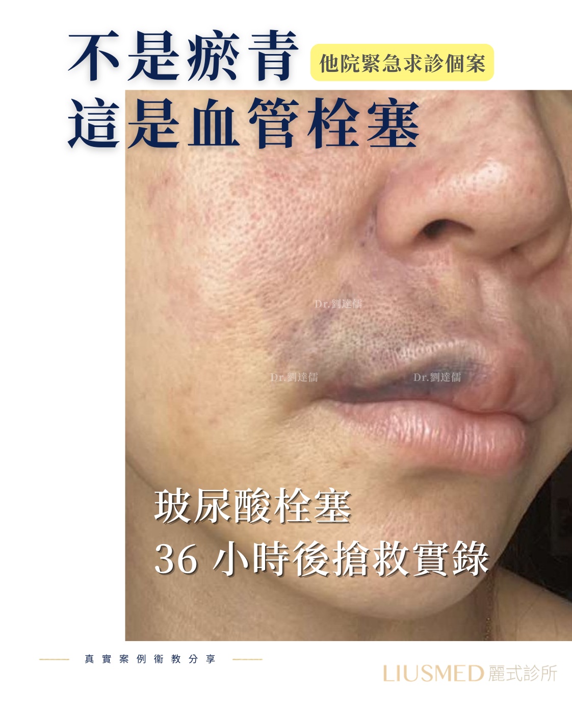
The second sign is a change in skin colour. Tissue downstream of a blocked vessel blanches first, then develops a mottled, marbled pattern, then turns dusky or greyish. The change follows the territory that vessel supplies, rather than staying around the needle entry point.
The third involves the eye. When injection has been done at the glabella, nasal dorsum, nose, tear trough or temple, sudden eye pain, a missing patch of vision, blurring or general dimming is the most time-critical presentation of all. That one needs emergency care immediately, not an appointment the following morning.
For any of these three, go now: eye symptoms are measured in minutes, and for the skin the earlier the better. The mechanism and why early treatment changes the outcome so much is set out in vascular occlusion: mechanism and emergency management, and our own emergency pathway is described under vascular occlusion rescue.
Because this stage turns on whether the injector recognises the pattern in the moment and has something on hand to act with, who holds the syringe matters considerably more than which brand of HA is in it.
Weeks to months later: the swelling that arrives out of nowhere
There is another pattern. Everything goes well, and then a month, three months, sometimes a year later you wake up to find the treated area swollen, tender to press, firmer than it was.
That is usually not filler "coming back out". Clinically it maps onto two things.
The first is a delayed inflammatory reaction. At some point the immune system starts responding to the material. The trigger can be a viral illness, dental work, a vaccination, or nothing identifiable at all. The typical picture is sudden onset, a distribution that matches where filler was placed, and a tendency to recur. How to tell it apart from ordinary post-procedure swelling is covered in swelling versus delayed inflammatory reaction.
The second is biofilm: bacteria living in a protective matrix on the surface of the material. It doesn't behave like an acute infection with heat and pus. It shows up as swelling, tenderness and firmness that keep coming back. The difficulty is that antibiotics often settle it and then it returns, for reasons explained in biofilm and recurrent filler swelling and why antibiotics alone fail.
The usual sequence is to control the inflammation medically first, then discuss what to do about the material once the acute phase has passed. What I want to flag is that the acute phase passing is not the end of the story. While the material is still in place, the same area can flare again. Results vary between individuals; the way to lower the chance of another flare is to deal with the source, not simply to wait for it to settle.
Key Insight: Swelling months later doesn't mean the original injection was botched, and it doesn't mean everything has to come out tomorrow. Separating inflammatory reaction from biofilm is what gets the treatment order right.
A year on: migration, encapsulation, and what ultrasound actually shows
Clear the earlier stages and a different set of problems takes over.
Migration is the most common. HA doesn't necessarily stay where it was placed. It travels along tissue planes, helped along by years of repeated muscle movement. Nasal filler drifting to the side of the nose, cheek filler descending toward the nasolabial fold, tear-trough filler settling into a visible ridge are all familiar. The routes it takes depend on which anatomical plane it was placed in, described in how fillers migrate.
Encapsulation is the second. The body walls long-standing foreign material off behind a layer of fibrous tissue. That changes two things: hyaluronidase can no longer reach the gel properly, and the nodule changes character under the fingers, from a soft pocket to a hard lump. Why dissolving stops working once a capsule has formed is set out in encapsulation and failed dissolution and seven reasons hyaluronidase fails. Hyaluronidase remains a useful tool, but whether and when to use it is decided after in-person assessment by a physician, not assumed because it worked for someone else.
By this stage, palpation can no longer carry the decision. Ultrasound answers three questions fingers can't: how much remains, which layer it occupies, and whether a capsule has formed around it. Those three answers are what decide between a medical dissolution approach and physical extraction. How the images are read is covered in ultrasound identification of fillers.
Our approach has always been to see it before touching it. Once position and plane are confirmed, the material is removed through a single pinhole under ultrasound guidance, working around vessels and nerves and disturbing as little healthy tissue as possible. That is the core of our filler revision service. How completely material can be retrieved varies from case to case, but the principle is straightforward: what you can see, you can clear properly.
Frequently Asked Questions
How long until HA filler has settled?
Swelling generally subsides and the shape stabilises at around two weeks. For a proper assessment of the final result, wait until the one-month mark. Mild asymmetry during that window often still changes, so there is no need to rush into a top-up.
I can feel a hard lump. Does that mean it failed?
In the first fortnight, firmness is usually oedema plus filler that hasn't integrated yet, and it softens. If it is still there past a month, has a distinct edge, or is present on one side only, that is worth imaging rather than continuing to wait.
Can HA be dissolved completely?
Whether hyaluronidase works is not decided by how long ago the filler went in; it still acts on HA that has been there for years. When dissolving stalls, the reason is usually a capsule around the gel, dense cross-linking, or something other than HA mixed into the injection, so the enzyme struggles to reach the gel and the result falls short of what people expect. How much can realistically be dissolved is decided from what the ultrasound shows, and outcomes vary between individuals.
It has been several years. Is it too late to do anything?
No, though the assessment shifts. With time, what matters is where the material is now, whether it has migrated, and how much fibrous tissue has formed around it. Those factors change which approach makes sense. They don't put the problem out of reach.
Finding out what is actually in your face
An imaging assessment is worth doing if any of these apply: you've had HA filler and aren't sure how much is left, you can feel a lump or see a contour that isn't right, you've had one episode of swelling months afterwards, or you were told it had all absorbed and you don't believe it.
At Filler Revision Clinic, Dr. Ta-Ju Liu scans the whole area with ultrasound first, confirms where the material is, how much remains and whether it has been encapsulated, and then talks through the options with you. Seeing it is what makes handling it safely possible.
For an initial online assessment and appointments, use contact us. International patients can share their current situation and treatment history remotely before travelling.
Written and reviewed by Dr. Ta-Ju Liu. This article is medical education and does not replace an in-person consultation. Individual situations should be assessed by a physician, and treatment outcomes vary between individuals.