"Doctor, was I given the blue vial or the black one?"
I have heard that question more often in the past few weeks than in the previous six months combined. The harder part is what usually follows — most people realise they do not know where to even look it up.
The reason is not mysterious. What patients see on social media is "blue vial, black vial." What clinic websites publish is "small-molecule, large-molecule." What appears on the consent form and the packaging is Juvelook or Juvelook Volume (Lenisna in some markets and records). Three vocabularies pointing at the same two products, and almost no page maps them onto one another.
So this happens: someone brings me quotes from two clinics and asks which is the better deal, and after all the comparing, the two quotes are for the same material. Or someone has reassured themselves that "mine was the skin booster type, so it should be fine" — and then describes an injection pattern that does not look like a skin booster at all.
What this article does is simple. It lays out the numbers for both vials, explains what that difference actually causes, and describes why, once a lump has formed, the plane it sits in matters more than the vial.
The names you have heard are two products from one line
Start by aligning the vocabulary. This is the premise for everything else, and it is the piece that is currently missing.
← Swipe to see more →
| What you may have heard | Which product it actually is |
|---|---|
| Blue vial, small-molecule, skin booster type | Juvelook, 50mg per vial |
| Black vial, large-molecule, volumising type | Juvelook Volume, 200mg per vial |
| Lenisna, "the 200mg one" | The same as above — the black vial |
That last row is the important one. Lenisna and Juvelook Volume are the same product, circulating under two names in both the literature and the market. A 2025 case report in the Journal of Cosmetic Dermatology writes it directly as "Juvelook Volume (Lenisna, VAIM Inc., Seoul, Korea)," placing both names inside a single parenthesis.
One honest caveat. A common claim is that "Juvelook Volume is the Korean name and Lenisna is the international one," but I checked the manufacturer's own product pages and found no such mapping stated anywhere. The accurate version is narrower: one product, two names in circulation. Which name belongs to which market is not something the manufacturer spells out.
Taiwan's licence record adds one more detail. The Juvelook licence (衛部醫器輸字第037021號) once listed a specification called "Lenisna 200", and that specification was cancelled on 13 August 2025. The instructions for use currently approved list two products, Juvelook 50 mg and Juvelook Volume 200 mg, so the black vial, Juvelook Volume, is still on the licence. The licence does not give a reason for the cancellation, so I will not guess whether it was a renaming or something else. In practical terms, under Taiwan's current licence the 200 mg vial is named Juvelook Volume; if your records say Lenisna, simply bring the product name and the injection date to your assessment.
Worth adding: "blue vial" and "black vial" are not the manufacturer's official naming either. They are nicknames that grew out of distribution. That is precisely why you rarely find those two words on a clinic website — clinics publish the specification, patients search the cap colour.
Key point: Before comparing what two clinics told you, confirm they are describing the same product. If the names do not line up, none of the comparison that follows means anything.
As for why the Chinese nickname "youth needle" points at several different materials at once, I unpack that in Juvelook lumps and nodules: what to do and will not repeat it here.
What actually differs: the numbers, laid out
There is no shortage of comparison articles, but almost none of them state where their numbers came from. That matters — for one single specification I found three mutually contradictory sets of figures. So below, each row comes with its evidence tier.
← Swipe to see more →
| Blue vial Juvelook | Black vial Juvelook Volume / Lenisna | |
|---|---|---|
| Total per vial | 50 mg | 200 mg |
| PDLLA | 42.5 mg | 170 mg |
| Non-cross-linked hyaluronic acid | 7.5 mg | 30 mg |
| Ratio, once converted | 85:15 | 85:15 |
These four figures carry two independent layers of support. The manufacturer's official product pages list "PLA (Poly-D,L-Lactic Acid) 42.5mg + HA (Sodium Hyaluronate) 7.5mg / Vial" and the corresponding 170mg + 30mg. A peer-reviewed journal paper states the same composition verbatim in its materials and methods section and names the product. Of every specification I checked while putting this together, this is one of the few with that kind of double sourcing.
The instructions for use approved in Taiwan reconstitute the blue vial with 0.75 mL of sterile water and the black vial with 3 mL: four times the product in four times the water, so the concentration per mL is the same. How concentrated each injection actually is depends on how the clinic dilutes it.
The instructions for use themselves say that an adequate dilution ratio, together with an appropriate dose, can reduce the chance of nodules after injection; the same document also warns against injecting into the superficial skin, to avoid papules or nodules. Published practice can differ widely from the label: one study of Juvelook in the nasolabial folds diluted each vial with 6 mL of saline. One point is often missed: if the whole vial goes in, the PDLLA that reaches your face is still 42.5 mg or 170 mg, however much water was added. Dilution changes how concentrated and how spread out each injection is, not the total.
A word on the ingredient itself. PDLLA (poly-D,L-lactic acid) is a resorbable polymer microsphere. Its job is not to fill a hollow directly but to remain in the tissue and stimulate your own collagen over time. The 15% that is hyaluronic acid is non-cross-linked, mainly providing even dispersion and immediate volume on the day, and it metabolises quickly.
While we are here: you will occasionally see Juvelook's ingredient written as PLLA, which belongs to a different material (Sculptra). You will also see PDLLA confused with PDO (polydioxanone), the material used in thread lifts. Three similar-looking abbreviations, three different materials, and three different approaches when something goes wrong.
So what does an identical ratio mean?
It means the blue vial is not a diluted hyaluronic acid product. It is equally a PDLLA-based collagen stimulator; there is simply less of it. The difference lies in total load (the brochure also lists different particle sizes and planes) — not in whether it stimulates collagen.
There is also a step that often gets skipped: the black vial carries four times the PDLLA of the blue. Vial for vial, the quantity of material sitting in your tissue and driving collagen production is on a different order.
Why "the blue vial is just a skin booster, so it cannot cause nodules" does not hold up
This is the sentence I most wanted to address here.
It circulates widely and it sounds reasonable: a skin booster goes in shallow, thin, and in small amounts, so how could it form a lump? I hear patients use this line to reassure themselves in clinic.
But if the ratio is identical, the premise collapses. There are 42.5mg of PDLLA in the blue vial. That is a real quantity of collagen-stimulating material, and it does not stop stimulating collagen because the treatment is marketed as a booster.
The more useful statement is this: a finer particle does not mean no collagen stimulation. The blue vial is often diluted and spread across the face as a skin-quality treatment, so it ends up shallow mainly because of how it is injected.
And more superficially can be the harder problem. The same cluster of material sitting deep may only be palpable; sitting in the superficial dermis, it may be visible — raised points on the skin surface, sometimes with a difference in how the area catches the light. Patients describe it as "grainy," or "little bumps I can feel."
I looked through the pages currently ranking on this question, and the handling is remarkably consistent: either the point is skirted, or it is closed with some version of "as long as you choose an experienced injector, there is no need to worry." That answer avoids the actual question. Patients are not asking whether to choose a good injector; they are asking whether the risk profile of these two vials is the same.
My answer: both can. What forms and where it sits depends mainly on the plane it was placed in, and so does the way it has to be handled.
On particle size, no two sources agree
Here I have to be straightforward about something I could not resolve.
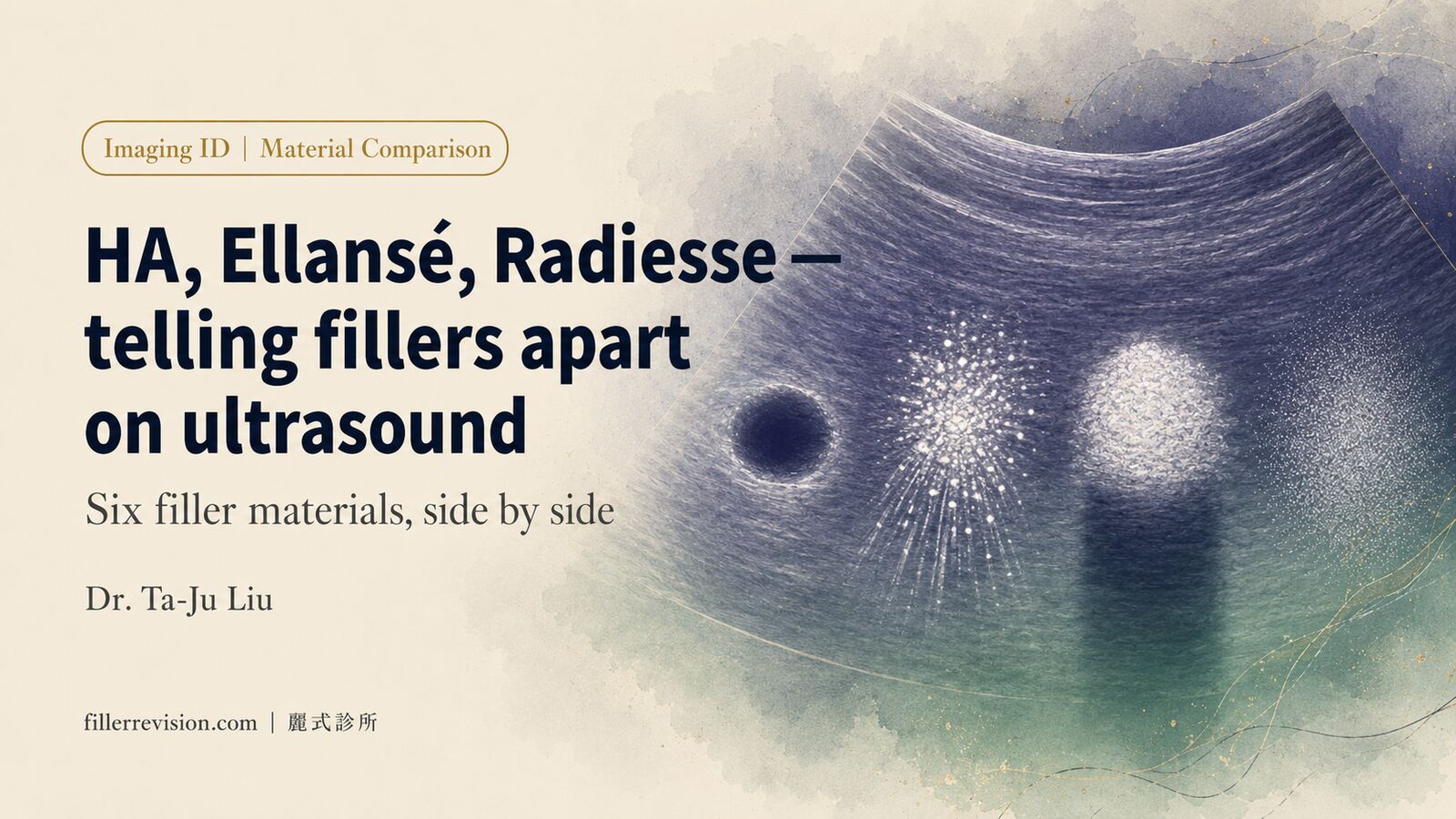
Particle size is the centrepiece of many comparison articles. When I laid out every source I could find, they contradicted each other:
← Swipe to see more →
| Source | Blue vial | Black vial |
|---|---|---|
| Manufacturer product brochure | 20–40 μm | 40–80 μm |
| 2025 case report | — | 40–60 μm |
| 2024 literature review, figure caption | 30–70 μm | — |
| Some distributor and clinic pages | 10–40 μm | 40–60 μm |
| Taiwan-approved instructions for use (licence 衛部醫器輸字第037021號) | 10–100 μm | 10–100 μm |
Five sets of figures, and four different accounts of the blue vial alone. Almost none of the consumer-facing pages state where their number came from.
So this is as far as I can honestly go: most commercial sources put the black vial's particles larger than the blue vial's, but the instructions for use approved in Taiwan list the same 10–100 μm range for both, so even that direction is not settled. The precise micron figure has no reliable consensus, and where you see a single set of numbers presented as settled, I would suggest asking for the source.
This is not pedantry. Many comparison pieces use particle size to infer which vial ends up in which plane, yet the instructions for use approved in Taiwan give both products the same particle range and the same injection site. Where the material finally sits depends on how it is diluted, which needle is used and how deep it goes. That is mainly technique, not the vial. Risk reasoning built on an unsourced number produces an equally unreliable conclusion.
The difference is not whether it stimulates, it is which plane it stops in
If I had to compress what separates one lump from another into one sentence:
Key point: A nodule is not a single disease. It is a question of location. Arguing over whether the product is good gets nowhere; establish which plane it is in and which type it is.
The manufacturer's brochure places the blue vial in the dermis and the black vial in the subcutaneous layer, rather than the "deep dermis" some descriptions use. The case report cited above also specifies that the blue vial is used in the upper dermis of the face.
The instructions for use approved in Taiwan do not make that split, though. Both products are listed with the same injection site, the dermis; the label allows use only in the dermis and subcutaneous tissue, and the approved indication is filling depressions of the nasolabial folds in adults aged 19 and over. The blue-for-dermis, black-for-subcutaneous distinction comes from the manufacturer's brochure and from clinical practice, not from the approved label.
Different planes produce three practical differences.
First, visible versus palpable. Material sitting superficially is usually something the patient sees; material in the subcutaneous plane is usually something they feel. This is why some people insist they have no lumps while a magnified photograph shows clear surface irregularity.
Second, the difficulty of dealing with it. Superficial material sits close to the surface, so removal has to account for surface smoothness and scarring. Deeper material involves surrounding vessels, nerves and other structures, which have to be visualised before deciding whether it can be touched at all.
Third, the timeline. I cover this in the pillar article, but briefly: swelling in the first days, non-inflammatory material aggregation at around three weeks, and anything surfacing months or a year or two later leaning towards a delayed reaction.
Can a mesotherapy injector gun even deliver it?
This section comes from clinic practice, and I have not seen it addressed in any patient-facing material.
People often ask me: "If it is injected by machine, does that avoid nodules?" The machine they mean is usually a mesotherapy injector gun.
The first part of the answer matters most: the needle on those guns is very fine, and this material may not pass through it reliably. Even when it does, the amount actually entering the skin may be small.
That is not a criticism of the device. The point is that when we compare "machine injection causes fewer nodules," we may not be comparing like with like. If less material entered the tissue to begin with, fewer nodules is the expected result — but that reflects dose, not the safety of the delivery method.
As for what the gun genuinely contributes: it addresses one variable, making depth and dose per pass more consistent, which does remove one failure mode — too much in a single point, unevenly distributed. That is worth something. But it does not change the total load.
In other words, the machine lowers that one variable. It does not move overall risk down a tier.
And one closing note: for a lump that has already formed, whether it was placed by machine or by hand does not change which plane it is sitting in now. Relitigating the injection technique contributes little to what happens next.
Shallow and deep placement produce different lumps
Pulling the previous sections together, this is roughly how problems present depending on the plane:
← Swipe to see more →
| Shallow pattern (how the blue vial is usually used) | Deep pattern (how the black vial is usually used) | |
|---|---|---|
| How patients describe it | Grainy, small bumps, visible | One firm area, deeper, felt rather than seen |
| Common sites | Forehead, periorbital area, full-face skin-quality zones | Tear trough, mid-cheek, nasolabial folds, cheeks |
| What assessment focuses on | Depth, density of distribution, surface smoothness | Extent, relationship to nerves and vessels, state of surrounding tissue |
This is a tendency, not a clean boundary. Both patterns can appear on the same face, particularly in someone who has had multiple sessions in different areas over time. The sites in the table are where lumps turn up in clinic, not recommended or approved sites: the instructions for use approved in Taiwan list the forehead and the area around the eyes as contraindicated, and the only approved site is the nasolabial folds.
One point about thin-skinned areas. Some content currently recommends the blue vial for the tear trough, but I rarely see anyone discuss the black vial's risk in the under-eye region. The thinner the skin and the more limited the subcutaneous space, the more readily the same quantity of material becomes visible. That has nothing to do with product quality; it is anatomy.
The instructions for use approved in Taiwan list the glabella, forehead, nose and the area around the eyes (such as the under-eye ring, eyelids and crow's feet) as contraindicated sites, and warn that injection into a vessel can cause serious harm such as loss of vision.
Hyaluronidase only dissolves that 15%
This is the most misunderstood aspect of Juvelook, and the most common source of frustration I see in clinic.
Hyaluronidase breaks down hyaluronic acid. Hyaluronic acid is 15% of what is in the vial. The remaining 85% — the PDLLA, plus the collagen it has already stimulated — is outside the enzyme's reach.
So the typical course runs like this: hyaluronidase is injected, the lump shrinks somewhat, the patient believes it worked, and some weeks later it is back to where it was. Only part of it was ever dissolvable.
This is what semi-reversible means: not irreversible, but nowhere near "one injection and it is undone." I set out the mechanism more fully in Why hyaluronidase fails, including what repeated enzyme injections cost in their own right.
The vial difference shows up here too: with four times the PDLLA, the portion hyaluronidase cannot reach is on a different scale in the black vial.
If you want to compare reversibility across several next-generation stimulators, the reversibility comparison sets them side by side.
Already injected and trying to work out which one? Three clues
Many people start looking into this after the fact. Here is what can realistically be established, ordered from most to least reliable.
First, ask for the record. The consent form, receipt or chart usually carries the product name, and some clinics retain the vial label or lot number. You are entitled to ask what was injected into you — that is not being difficult, it is basic medical information. Packaging currently approved in Taiwan reads Juvelook or Juvelook Volume, with 50 mg or 200 mg; in other markets or other records, the 200 mg vial may appear as Lenisna.
Second, consider the site and the goal. If the aim was skin texture, pores and fine lines, spread over a wide area, it was most likely the blue vial. If the aim was lift, hollows or structural support, concentrated in the tear trough, mid-cheek or nasolabial folds, it was more likely the black vial.
Third, recall the technique. Wide-area, multi-point, device-assisted delivery points towards the blue vial; slow deposition through a needle or cannula at specific points points towards the black.
The third clue is the weakest and should be treated as circumstantial only. The first is the one that settles it.
Mixing the two, and stacking sessions
Plenty of people ask about this and few get a serious answer.
Using the two in different areas and different planes is a common enough pairing. What needs attention is not whether they can be combined, but total load and the interval between sessions.
I have seen accumulated quantities in clinic that were considerable — repeated top-ups over a short period, each time on the reasoning that "the result still is not obvious, let us add a little more." The difficulty is that PDLLA takes time to stimulate collagen, usually weeks to months before the change is apparent. Topping up before the previous session has fully expressed itself makes it easy to stack far more total material than intended.
By the time the collagen does appear, what appears is the sum of all of it.
Key point: "I cannot feel anything yet, let us add a bit more" is the single most dangerous line of reasoning with this material. It is slow by design. Not seeing a change does not mean nothing is happening.
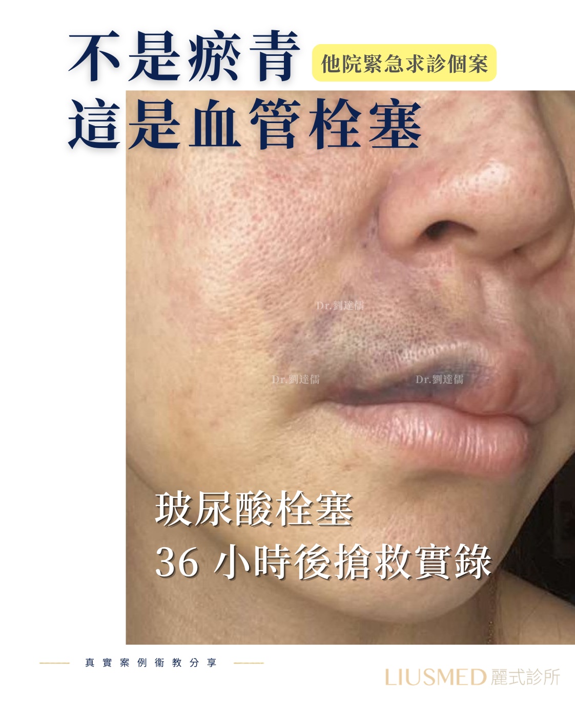
Both vials carry vascular risk. The instructions for use approved in Taiwan warn that use in a blood vessel can cause vascular occlusion and tissue necrosis, and that injection into a vessel can cause serious harm such as loss of vision. Any change in vision after an injection means going straight to an emergency department or eye doctor; pain that keeps getting worse or skin turning pale or mottled needs the injecting clinic or an emergency department now. What to expect when the material cannot be dissolved is covered in vascular occlusion from non-HA fillers.
When to act, and when to leave it alone
I will keep this short, because the full triage sits in the pillar article.
The principle: small nodules that are asymptomatic and do not affect appearance do not need urgent intervention — monitoring is enough. What warrants active assessment is inflammation, continued growth, an effect on appearance, or a lump that keeps returning despite repeated treatment.
Whichever vial caused it, my approach is the same: use ultrasound to establish position, depth and surrounding structures first, then decide whether it can be touched and how. Being able to see it is the precondition for discussing safe handling, and that is the basis of filler lump extraction.
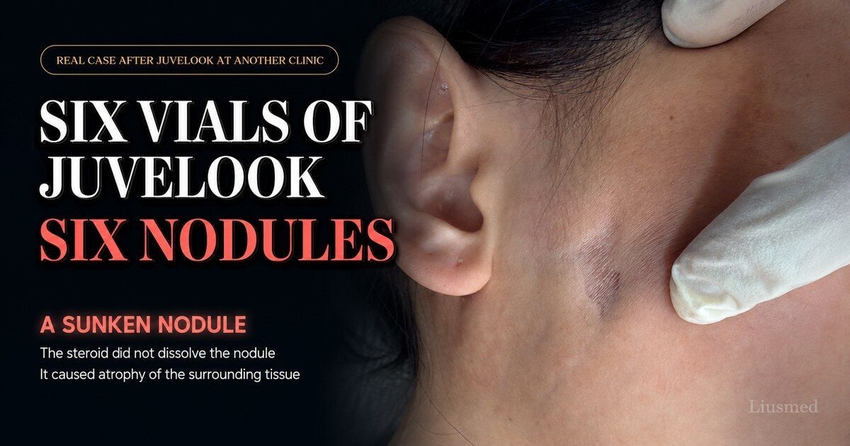
One thing to note: whether steroid has previously been injected into the lump materially changes the difficulty of everything afterwards. If your doctor has proposed injecting steroid into a lump, I would read the risks of steroid injection and tissue atrophy before deciding.
For the full nodule triage and treatment ladder, see Juvelook lumps and nodules: what to do. To orient yourself on which category you may fall into, the collagen stimulator condition overview is a reasonable starting point.
Frequently Asked Questions
Is Juvelook injected in Korea the same product as in my own country?
The specification belongs to the same product line, but the real difference is not the material — it is whether anyone can be reached afterwards. The recurring difficulty with treatment abroad is that lot number, dilution, injection plane and dose records are unavailable, so anyone treating a complication later is rebuilding the history from nothing. This is not a comment on any country's standards; it is a structural break in the follow-up chain. If you are treated overseas, record the product name, dose and sites before you leave.
How many vials do I need?
For the nasolabial folds, the instructions for use approved in Taiwan set limits of 1.2 mL per side per session and 2.4 mL per treatment session, with any further correction assessed only after 4 weeks. That depends on area, extent and individual factors, and there is no universal answer. I would reverse the question: confirm that the previous session has fully expressed itself before deciding whether to add more. PDLLA works gradually, and topping up before the result has arrived is the most common route to an excessive total that I see.
I have felt nothing for months. Was I sold something that does not work?
Not necessarily. This material is not designed to show an immediate result. The immediate plumpness comes from that 15% of hyaluronic acid, which disappears once it metabolises; the collagen itself takes considerably longer. That said, "nothing at all" may also reflect dose, plane or distribution, so it is worth returning for review — but not worth topping up while the reason is unknown.
Can the blue vial be used in the tear trough?
The instructions for use approved in Taiwan list the area around the eyes, including the under-eye ring and eyelids, as a contraindicated site, and the only approved site is the nasolabial folds. The under-eye area also has some of the thinnest skin on the face and limited subcutaneous space, which makes the same quantity of material most visible there. If it has already been injected there and a lump has formed, an in-person assessment with ultrasound is the next step.
I already have a lump. Can more product be layered over it?
I would advise against it. Adding material to an area that already contains an aggregate rarely smooths the surface, and it usually makes the quantity and the planes involved more complicated to address later. Getting the current situation properly assessed matters more than a quick correction.
A closing thought
Back to the opening question: which one were you given?
I hope this article at least makes that question answerable. The names line up, the figures have sources, and the planes are described clearly — so that when you ask your own doctor, you know what you are asking and can follow the answer.
As for "which vial is safer," my answer is that it is not a good question. What actually determines risk is how much went in, which plane it went into, what kind of skin it went under, and whether more was added before the previous session had shown its result. The material is one variable among several, and not the largest one.
If you already have a lump, or are unsure whether your situation needs treating, you are welcome to arrange an assessment. I will use ultrasound to establish position and depth first, then discuss with you whether it should be touched, and how.
References
- Seo SB, Park H, Jo JY, et al. Skin rejuvenation effect of the combined PDLLA and non cross-linked hyaluronic acid: A preliminary study. J Cosmet Dermatol. 2024;23(3):794-802. PMID: 37969055
- Seo SB, Wan J, Yi KH. Energy-Based Device Management of Nodular Reaction Following Poly-D, L-Lactic Acid Injection for Tear Trough Rejuvenation. J Cosmet Dermatol. 2025;24(1):e16575. PMID: 39283001
- Lee KWA, Chan LKW, Lee AWK, et al. Poly-d,l-lactic Acid (PDLLA) Application in Dermatology: A Literature Review. Polymers (Basel). 2024;16(18):2583. PMID: 39339047
- VAIM Co., Ltd. Manufacturer product specifications (Juvelook / Lenisna)
- Taiwan Food and Drug Administration. "VAIM" Juvelook Soft Tissue Injectable Implant, approved Chinese instructions for use, licence 衛部醫器輸字第037021號 (TFDA approval stamp dated 2025-09-05): product specifications, intended use, directions (reconstitution and dosage), warnings 6, 12 and 13, potential adverse effect 4h, contraindication 6.
- Yi KH, Rosellini I, Min JLJ, Kim HS. A Clinical Study on the Efficacy and Safety of Injectable PDLLA for Nasolabial Fold Correction: Juvelook treatment for Nasolabial fold. J Craniofac Surg. 2026 (online ahead of print). doi:10.1097/SCS.0000000000012819. PMID: 42479568
- Taiwan Food and Drug Administration. Medical device licence open data (dataset 68), licence 衛部醫器輸字第037021號: specification field recording that the "Lenisna 200" specification was cancelled on 2025-08-13 (accessed 2026-10-21).