
"Six months of steroid injections and the lump is still there. My doctor says there's nothing else they can do — is that really true?" At FILLER REVISION, we hear this from biostimulator patients every week. This is the defining frustration of biostimulator complications. Poly-L-lactic acid (Sculptra), polycaprolactone (Ellansé), and calcium hydroxylapatite (Radiesse) cannot be dissolved by any enzyme — that's a fact. Published nodule incidence rates range from 1-10% depending on the product and technique. But "can't be dissolved" doesn't mean "can't be removed." These particles become embedded within newly-formed collagen matrix, creating a composite material that resists chemical dissolution — but responds to physical extraction.

Common Symptoms
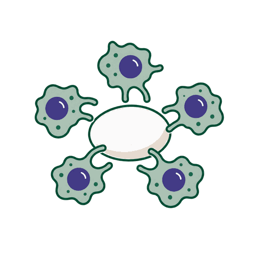
Particle Aggregation & Immune Response
If you've been going back every few weeks for steroid injections that shrink the lump temporarily but never make it go away, you're not failing treatment — the treatment is failing you. Here's why it never works long-term. Improper dilution, inadequate mixing, or injection into the wrong tissue plane causes particle aggregation—clusters of microspheres that trigger an exaggerated foreign body response. The body attempts to wall off these clusters with dense fibrous tissue, forming palpable nodules. Second, in susceptible individuals, the immune system mounts a granulomatous reaction—recruiting macrophages, giant cells, and inflammatory mediators that form an organized inflammatory mass around the particles. Unlike HA complications, these cannot be chemically dissolved; the particles persist within a collagen-fibrous matrix that only responds to physical intervention.
Why Traditional Treatments Fail
Why Conservative Treatments Fail
Since biostimulators are not hyaluronic acid, enzymatic dissolution has zero effect. Intralesional steroid injections can temporarily reduce inflammation and nodule size, but they do not remove the foreign body trigger—leading to recurrence once treatment is stopped. Prolonged steroid use carries significant risks: fat atrophy causing visible skin depressions, skin thinning, telangiectasia, and hypopigmentation. Other injectable agents can soften early-stage inflammatory nodules but have limited efficacy against mature, encapsulated nodules where dense scar tissue has already formed around the particle clusters. The fundamental problem remains: without physically removing the foreign particles, the body's immune response will continue indefinitely.
“At FILLER REVISION, biostimulator patients are often the most exhausted — they've been through months of steroid cycles, watching the lump shrink and swell like clockwork. The moment I show them the ultrasound image of the particle cluster inside the capsule, they immediately understand: no injection will ever dissolve this. It needs to come out. And once it does, the cycle is finally over.”
Dr. LiuWhen Nothing Can Dissolve It, Physical Extraction Is the Direct Route
Ultrasound-Guided Pinhole Micro-Extraction
Biostimulator particles — PLLA, PCL, CaHA — are immune to every enzyme and medication available. Physical extraction addresses them directly. But the critical challenge isn't just removing the foreign particles; it's removing them while preserving the patient's own fat and collagen that surround them.
Months of Steroids Got You Nowhere — Here's Why
At FILLER REVISION, patients arrive after 3-6 months of steroid injections that shrank and swelled the same lump over and over. No chemical on earth dissolves PLLA, PCL, or CaHA — steroids only temporarily quiet the immune response while the particles sit untouched inside.
Precision Extraction Separates Foreign From Natural
The hardest part of biostimulator revision isn't removing the particles — it's removing them without destroying the patient's own healthy fat and collagen that surround them. This is where a revision specialist's daily experience makes the difference.
End the Steroid Damage Before It Gets Worse
Each steroid cycle carries its own risks: fat atrophy, skin thinning, visible depressions. Some patients arrive at FILLER REVISION with steroid damage worse than the original nodule. Physical extraction ends both problems at once.
Pinhole Micro-Extraction
Our approach targets the root cause: physical removal of the foreign particle clusters. Using ultrasound to precisely locate each nodule, we employ micro-dissection to separate the nodule from surrounding healthy tissue, then mechanically fragment the compacted material into extractable pieces. The fragments are aspirated through a pinhole entry point. This addresses the source, as much as can be safely removed, without the tissue damage, scarring, or fat atrophy associated with open surgery or repeated steroid injections.
Ultrasound Localization
Micro-Dissection
Mechanical Fragmentation
Aspiration & Verification
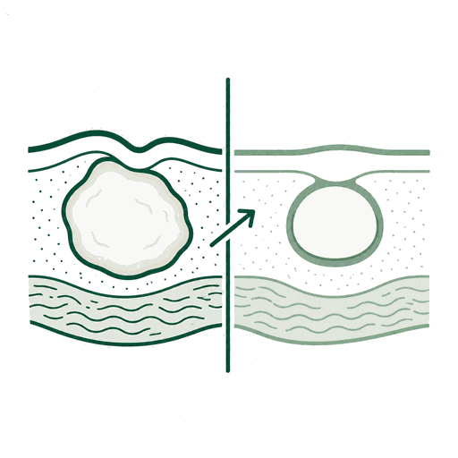
Before & After Results
View real patient results for this condition, including ultrasound imaging before and after extraction.
View All Case ResultsWatch: Real Removal Case
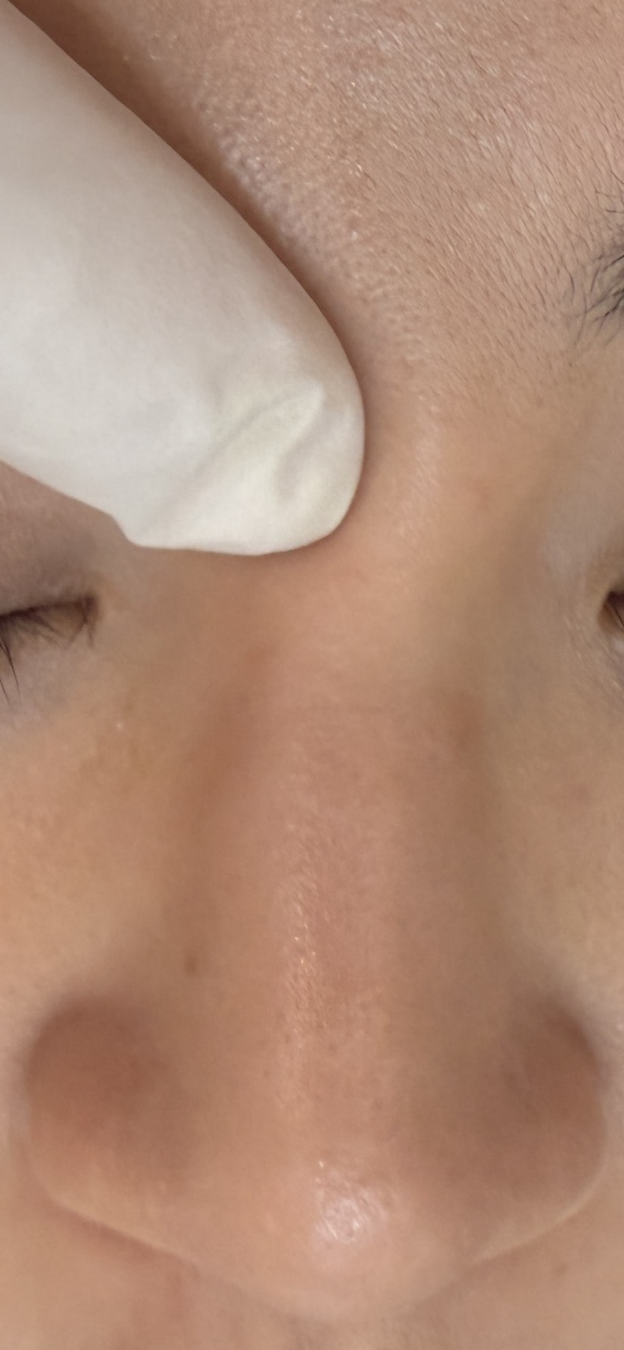
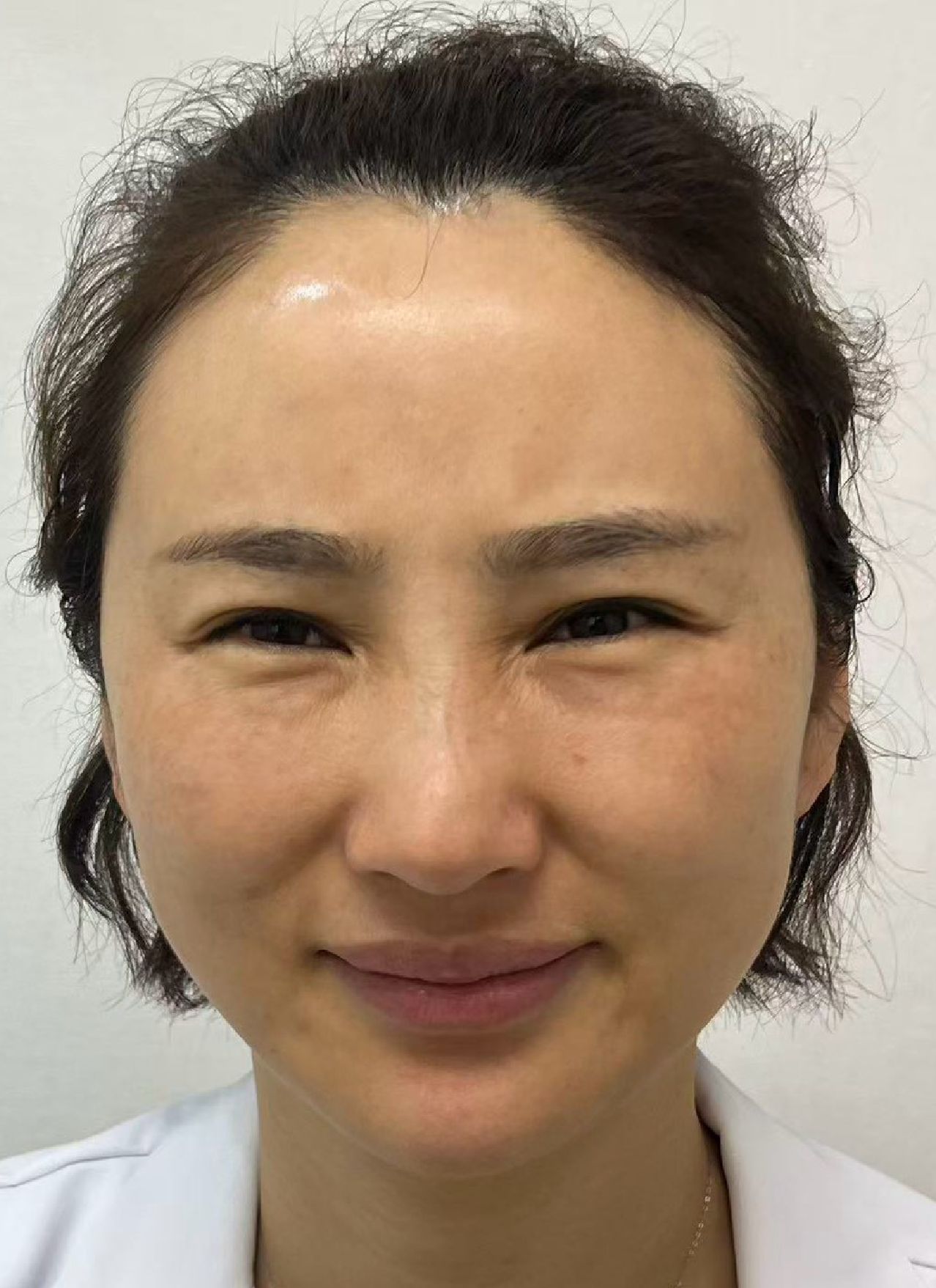
Under-eye lumps from Sculptra & Ellansé removed through a single pinhole under ultrasound guidance — no open surgery, and the pinhole is usually barely visible once healed.
Available in English · 中文 · 日本語 · 한국어
Common Questions
Small subclinical micronodules may soften with diligent massage, but observation should be limited to about three months. However, visible or palpable nodules—especially those that have developed a fibrous capsule—rarely resolve spontaneously. Clinical evidence shows that mature nodules persist indefinitely without physical intervention. Early detection with ultrasound allows simpler treatment before the capsule fully matures.
Ellanse (polycaprolactone microspheres in carboxymethylcellulose gel) tends to form denser, more cohesive nodules than Sculptra (poly-L-lactic acid). Both require physical extraction, but Ellanse nodules typically need more mechanical fragmentation due to their material properties. Both can be treated with our ultrasound-guided technique; as with any extraction, 100% removal can't be guaranteed.
No open surgery is needed. We do not perform open surgery. Our pinhole micro-extraction technique works through a needle-sized entry point of about 2 mm; the pinhole heals in about two weeks and is usually barely visible afterwards. When nodules are large or deep, we treat them zone by zone over separate sessions, still through pinholes of about 2 mm, with no incision.
Once a nodule is physically extracted, that specific lesion does not recur. However, if residual product remains elsewhere in the treatment zone, new nodules can potentially form at those sites. This is why we perform comprehensive ultrasound scanning of the entire treatment area—not just the symptomatic spot—to identify and address as many deposits as can be treated safely; depending on the extent, this may take more than one session.
Our technique preserves surrounding healthy fat and tissue. Minor contour irregularity is possible for larger nodules, but this is typically much less noticeable than the nodule itself. If significant volume loss occurs, it can be addressed with fat grafting or safe fillers after complete healing.
Nodules typically appear 3-12 months after injection, coinciding with the peak of the body's foreign body response. However, some cases present 1-2 years later, particularly when triggered by immune activation events. Regular ultrasound monitoring in the first year after biostimulator injection enables early detection and simpler treatment.
Steroids suppress the inflammatory component of the nodule—reducing swelling and tenderness—but they cannot dissolve the synthetic polymer particles that triggered the reaction. Once the steroid effect wears off, the immune system re-engages with the foreign body, and the nodule returns. Repeated steroid use also risks fat atrophy, creating visible depressions that can be worse than the original nodule.
Unfortunately, yes. This rebound pattern is common. Steroids suppress the inflammatory component temporarily, but the foreign body (PLLA particles) remains. When the steroid effect wears off, the immune system re-engages — often more aggressively than before. Each cycle also adds more fibrosis, making the nodule progressively harder. This is why we advocate for extraction rather than repeated steroid suppression.
Ellansé's carrier gel (carboxymethylcellulose) does absorb, within about 6–8 weeks, but the polycaprolactone microspheres and the collagen they've stimulated do not simply disappear. If you have a palpable nodule, waiting typically means the body continues to build fibrous tissue around the particles, making the nodule harder and potentially larger. Early intervention produces better outcomes with less tissue disruption.
This is exactly why revision-only practices like FILLER REVISION exist. Your injector specializes in placing products — they aren't trained or equipped for extraction. You need ultrasound imaging to see the nodule, micro-instruments to extract it, and a doctor who does this daily. It's a different specialty entirely.
Not sure what is going on? Write it down and ask.
Describe what was injected, when, and what you are seeing now. Dr. Liu replies in the thread with an educational explanation of what it might be, what needs to be told apart, and which signs mean you should be seen soon. Posting can be anonymous, and other patients with the same problem can read the answer too.
Replies are general health education, not a diagnosis, and do not replace an in-person examination.

Read next: how we actually handle it
Case-by-case decisions, written by Dr. Ta-Ju Liu
Ellansé Lumps: What to Do About Early vs Delayed Nodules
Ellansé lump — what to do? Early vs delayed nodules, the side effects that matter, and how an uneven face after extraction elsewhere can still be revised.
Sculptra Granulomas and Delayed-Onset Nodules: Why They Form, and How Removal Is Planned
Why PLLA granulomas and delayed-onset nodules appear months after Sculptra, which risk factors are traceable, and how ultrasound-guided removal is planned.
HArmonyCa Nodules: Hybrid HA + CaHA Lumps, Half-Reversible
HArmonyCa (HA + CaHA hybrid): what it is, its side effects and nodule rate, and why hyaluronidase only half-works — plus how to get the hard core out.
AestheFill Nodules & Lumps: PDLLA Treatment Ladder, Removal
AestheFill gone wrong? PDLLA lumps and nodules: the treatment ladder from massage and lavage to ultrasound-guided removal, and when to stop injecting.
Radiesse Calcium Lumps: Why Hyaluronidase Cannot Remove
Radiesse lumps, jaw widening, or calcified nodules that won't resolve? Ultrasound-guided extraction is a definitive option when no dissolver exists.
5-FU & Steroids Failed Your Sculptra Lump? The Alternative
Steroid or 5-FU rounds done and the lump persists? Why drugs fail collagen stimulator lumps, and how ultrasound-guided extraction resolves them.
References
- Park SK, et al. Hypopigmentation and subcutaneous fat, muscle atrophy after local corticosteroid injection. Korean J Anesthesiol. 2013;65(6 Suppl):S59-S61.
- Aguilera SB, et al. Successful Treatment of Calcium Hydroxylapatite Nodules With Intralesional 5-Fluorouracil, Dexamethasone, and Triamcinolone. J Drugs Dermatol. 2016;15(9):1142-1143.
- Lemperle G, et al. Foreign body granulomas after all injectable dermal fillers: part 2. Treatment options. Plast Reconstr Surg. 2009;123(6):1864-1873.
Related Real Cases
Documented ultrasound-guided extraction and rescue cases by Dr. Ta-Ju Liu.
The information on this website is for educational purposes only and does not constitute medical advice. Individual results may vary depending on personal conditions; actual outcomes cannot be guaranteed. All medical procedures carry potential risks and complications. Please consult a qualified physician before making any treatment decisions.
Featured Poster
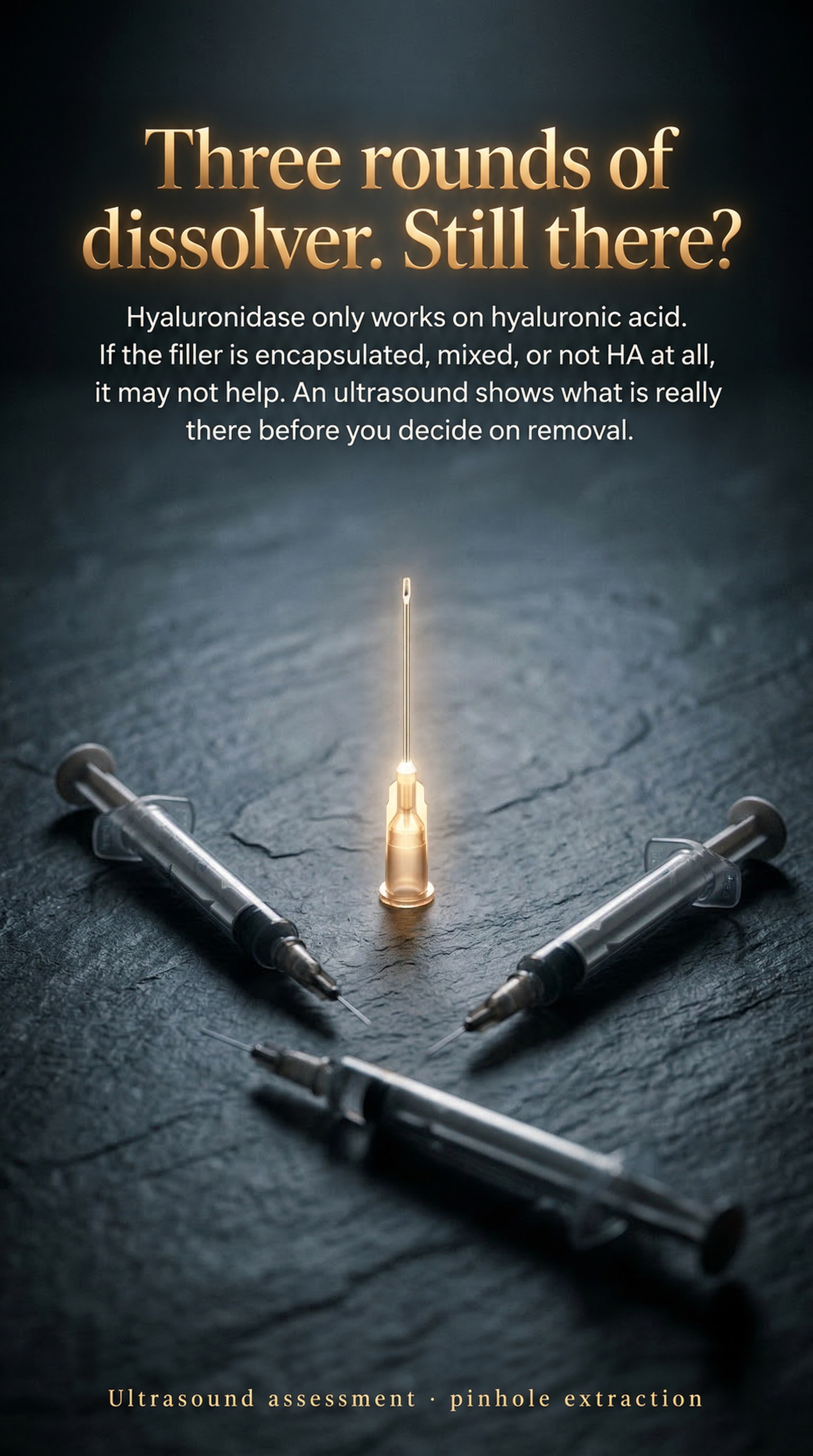
Three rounds of dissolver. Still there?
Hyaluronidase only works on hyaluronic acid. If the filler is encapsulated, mixed, or not HA at all, it may not help. An ultrasound shows what is really there before you decide on removal.

Dissolved — and it still hurts?
Only a little filler left, hyaluronidase several times, and still the headaches, facial pain, neck and shoulder aches. We look for what remains on ultrasound, and a board-certified neurologist on our team assesses the pain with us.