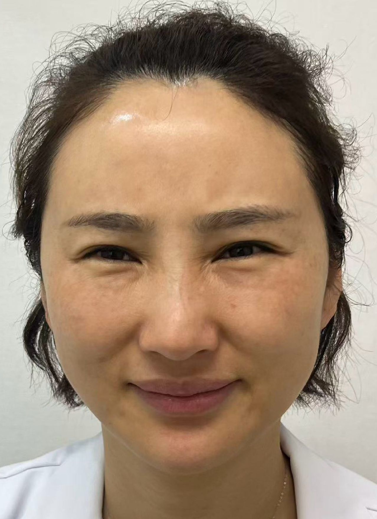
This is not a success story. It is here because it happens to form a comparison that could not have been arranged deliberately — and both sides of the comparison are on the same face.
The boundaries first. This is one patient's experience. The same material at a different volume, in a different plane, in a different person will behave differently. A single case cannot prove that Juvelook always causes nodules, and it cannot prove that it never does. All images are shared with the patient's consent and have been de-identified. Her injections were not performed at this clinic.
What she came in with
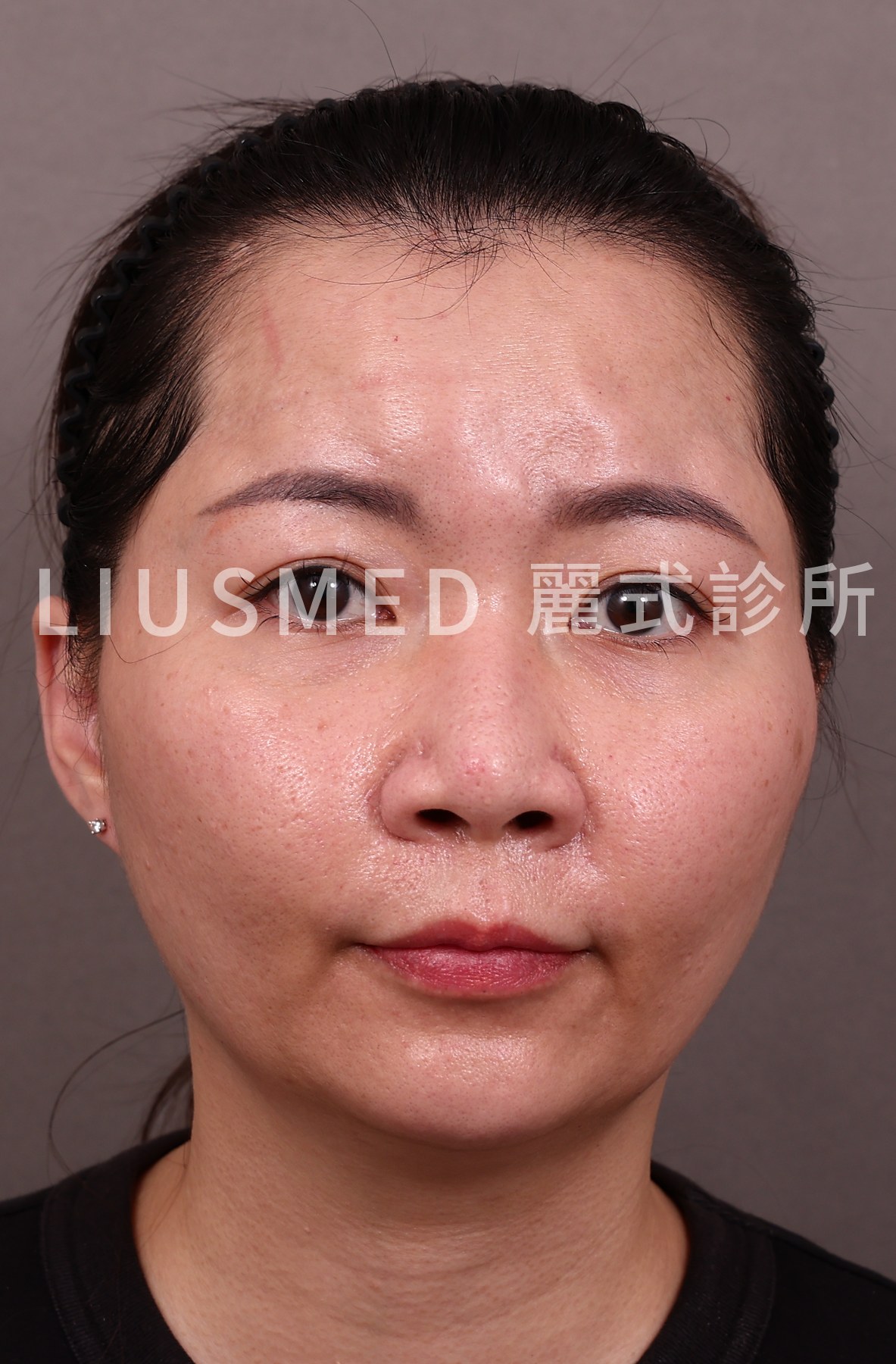
Six vials of Juvelook, given across three sessions over four months. Lumps had appeared in the tear troughs, the cheeks, the nasolabial folds, and from the temples down to just in front of the ears. The larger ones were roughly the size of a thumb tip.
That volume and that pacing are the starting point for everything that follows. Juvelook belongs to the collagen stimulator family — PDLLA microspheres combined with hyaluronic acid. The HA half can be dissolved; the PDLLA and the collagen it stimulates cannot. With this class of material, volume is not a parameter you get to adjust afterwards.
What ultrasound showed first
Before deciding whether to do anything, and what, we imaged every site with high-frequency ultrasound. Within one person, on one material, the pattern was not the same everywhere.
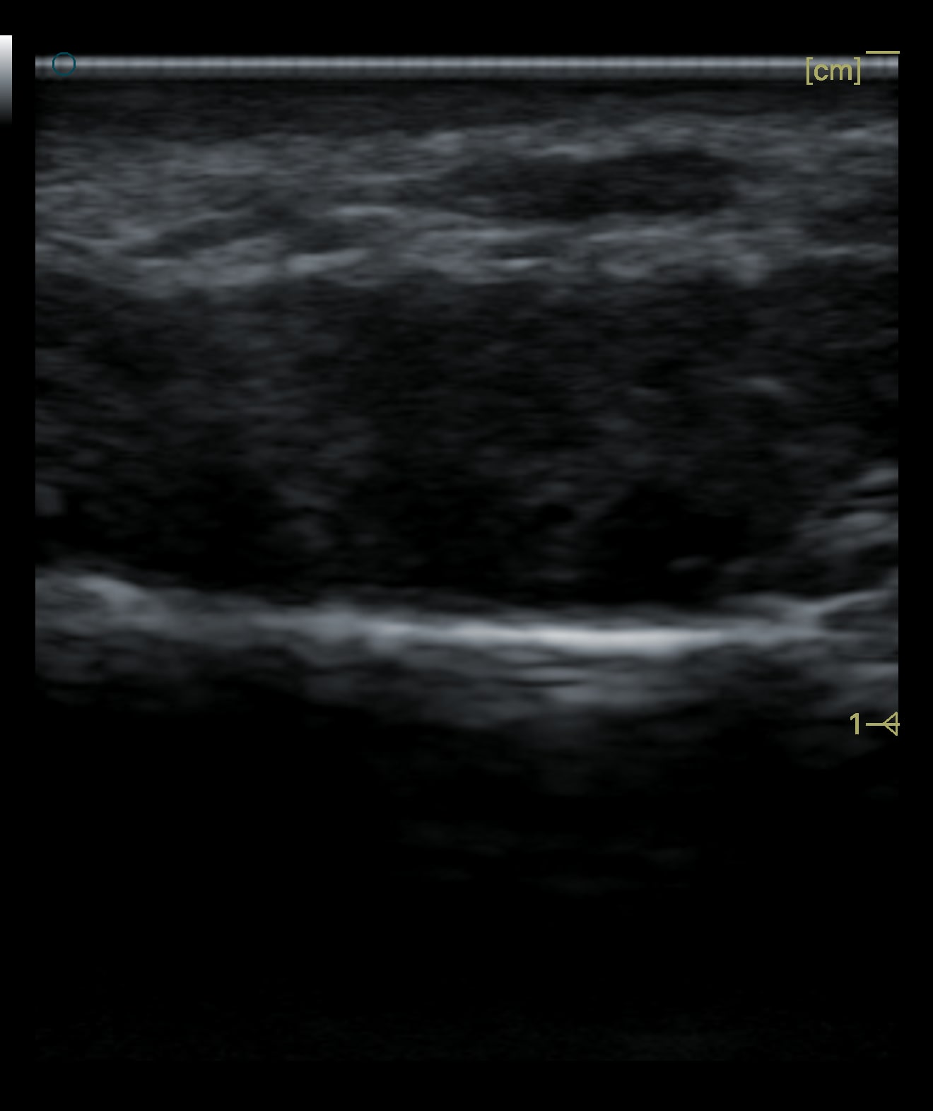
This site is one continuous mass. The border is reasonably distinct, and the tissue planes pushed aside beneath it are visible.
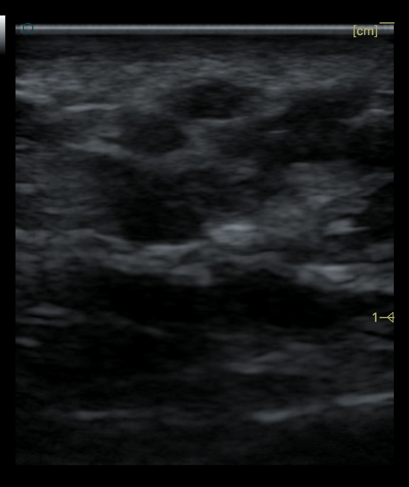
This one is different. The material is dispersed — individual deposits sitting within the tissue, with no border you could lift a whole mass out along.
That difference decides the difficulty directly. A confluent mass can be reduced layer by layer along its border. Scattered material has no border to follow; it has to be confirmed and addressed region by region under ultrasound, and how much can be reduced is inherently limited. This is why "can the lump be removed" has no yes-or-no answer. You have to see what shape it has taken first.
This was done in two sessions, not all at once
Worth stating early, because the photographs below span two dates.
The sites that had never received steroid — the under-eye area, meaning the tear troughs and mid-cheeks — were addressed first. The lateral cheeks followed three weeks later. That order was not arbitrary: the area that had received steroid had already lost a layer of tissue, so it was given time to settle, and it was worth seeing first how an undisturbed site looks after extraction before deciding how to approach the one that was not.
Taking every nodule out in a single sitting sounds more decisive, but the swelling stacks up and the reading gets worse with it — which area responded well and which did not becomes impossible to separate inside one recovery period. Staging is not inefficiency. It is what keeps the result readable.
The decision: her clinic recommended steroid across the whole face; she agreed to one site
After the lumps appeared she went back to the clinic that had injected her. The recommendation was steroid injection into the nodules — across the whole face.
She had read about it first, knew that steroid can cause tissue atrophy, and agreed to trial one site only: the area in front of the ear.
That decision turned out to be the most valuable part of this case. She had six nodules from the same material, the same technique and the same period. Five had never received steroid. One had. The single variable was that injection.
The site that received steroid: the nodule came out, the depression stayed
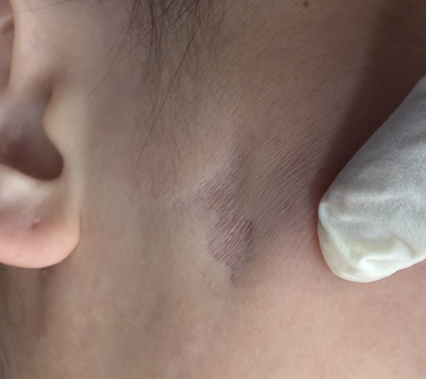
This is the site that received steroid, before extraction. What you see is not a lump standing up off the surface. It is a sunken, darker patch where the skin has thinned enough for the vessels underneath to show through. The nodule is deeper — palpable, but what shows first is the collapsed tissue around it.
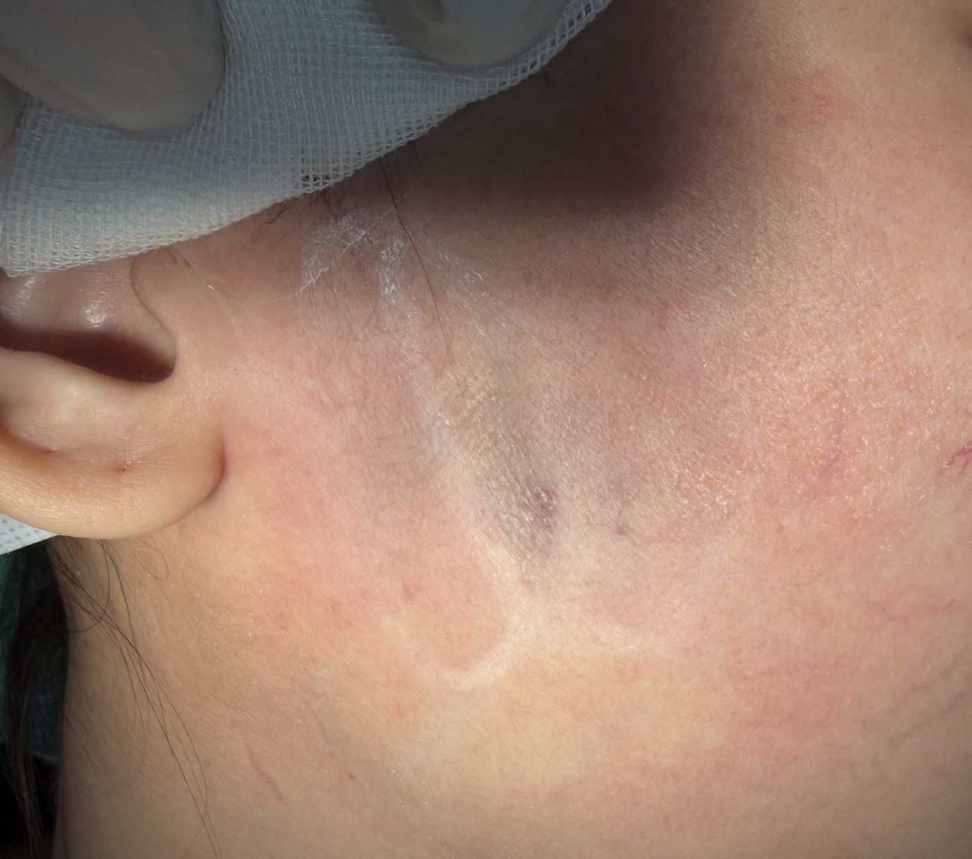
After extraction the nodule is gone. The depression is not.
This is the most important image here and the least flattering one. What it demonstrates: steroid suppresses inflammation and fibroblasts. It has nothing to act on where PDLLA already sits in the tissue. So the nodule stayed exactly as hard and as large as it was, and what was pressed down was the healthy fat and dermis beside it.
Put another way — that injection did not treat the problem. It treated the tissue next to the problem. And the tissue that atrophied is precisely what you need if you later have to address that nodule. Once the surrounding tissue has lost a layer, the working space shrinks with it, and the difficulty goes up rather than down.
The mechanism, which facial areas are most vulnerable, and what options remain once atrophy has occurred are covered in steroid injection and tissue atrophy.
The sites that never received steroid: marking and extraction
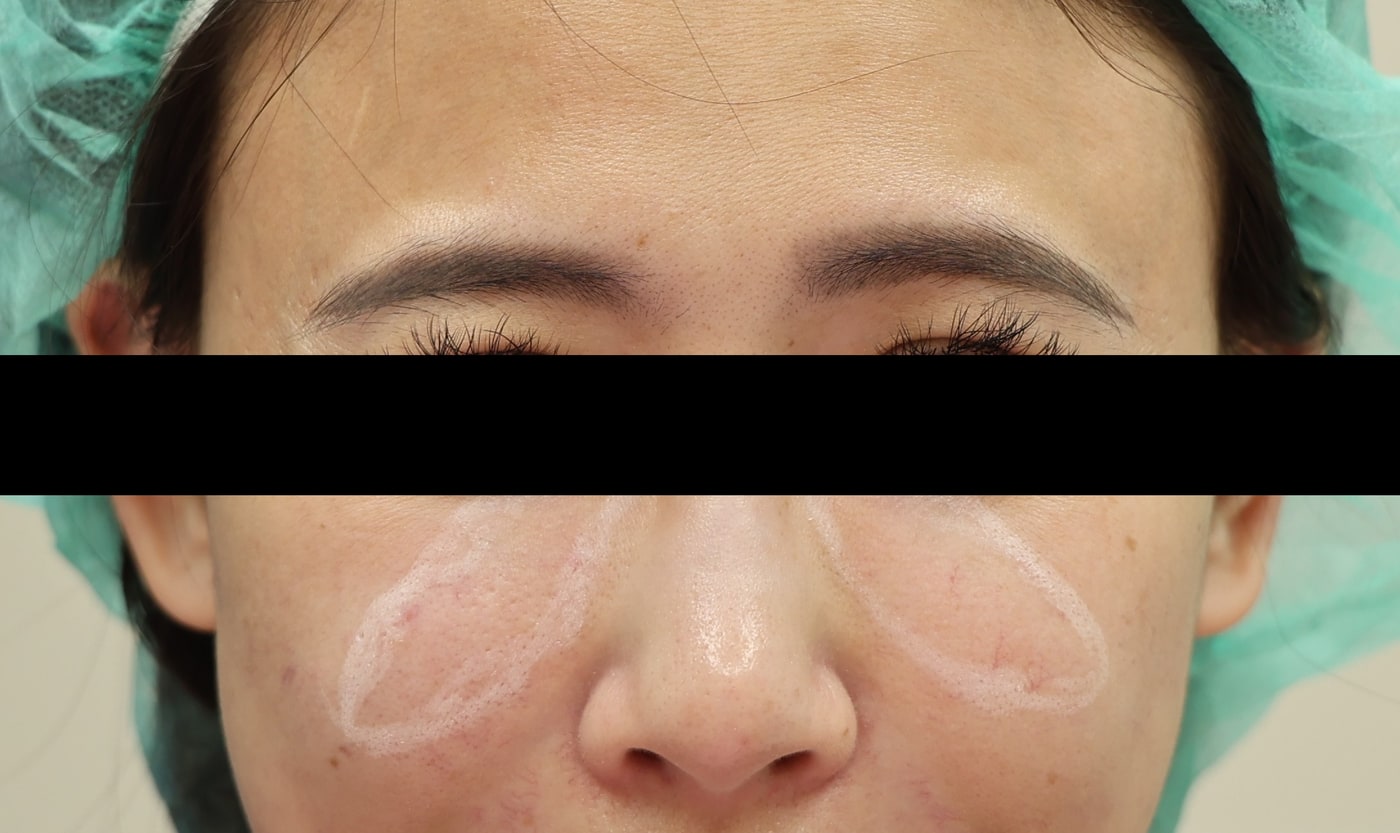
The tear trough and cheek areas were marked before the procedure. The marking is not an approximation — it is the actual distribution of the material, located on ultrasound and drawn onto the skin, and it determines where the pinhole entry goes and which direction to work in.
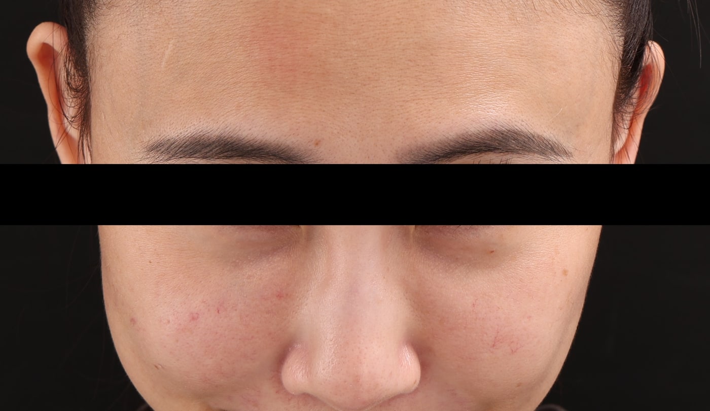
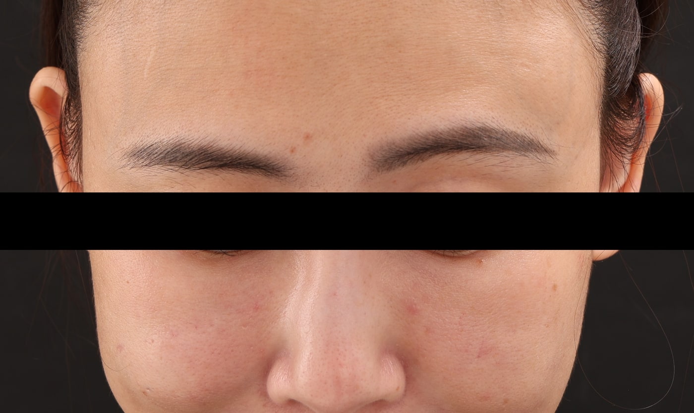
The sites that had never received steroid settled more evenly once the swelling phase passed. Recovery varies between people and between areas, and no general rule is being drawn from this here.
The extracted material
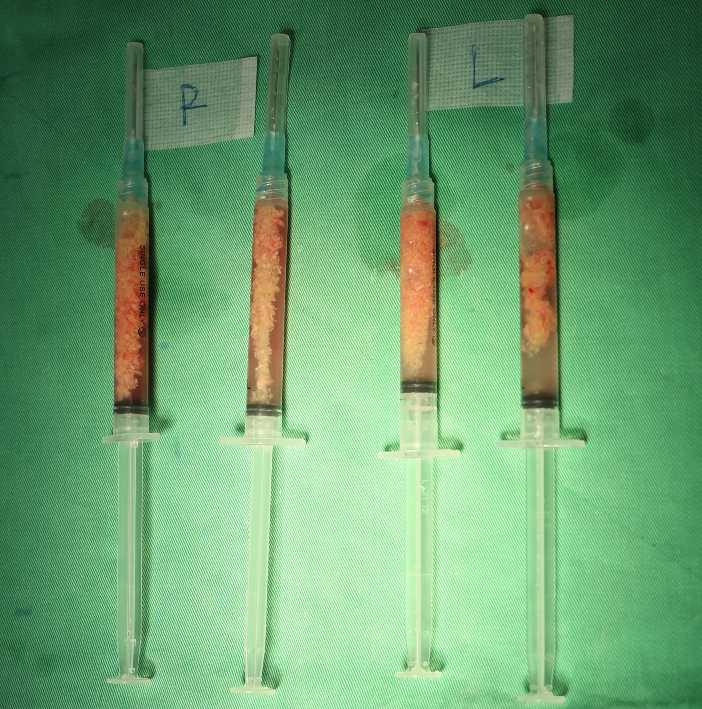
This is from the second session — the cheeks. Four syringes, right and left collected and labelled separately, holding particulate material mixed with tissue fluid.
The reason for including this photograph is simple. When the subject is filler removal, what came out is something you can look at directly, without needing anyone's adjectives.
What this case can and cannot support
What it supports. In this patient, for nodules caused by this material, steroid did not make the nodule smaller but did leave that site with less tissue. Same person, same period, same material — the variable is clean.
What it does not support. It does not support "Juvelook causes nodules." It does not support "steroid causes depressions" either — steroid still has its place in the right indication, and dose, concentration, depth and frequency all change the outcome. This is n=1.
One more thing worth saying plainly. The people who walk into a revision clinic are, by definition, the ones for whom things did not go smoothly. The people whose treatment went well never appear in my clinic, so my denominator is skewed by construction. I can describe honestly what I see. I cannot pretend that what I see is the whole picture.
What this procedure cost
The nodule in front of the ear was sitting against a nerve. Ultrasound guidance shows where a nerve runs and allows you to work around it; what it cannot change is that the material had grown there. After dissection, nerve function in that area was affected, and there was a period of facial asymmetry.
It has been improving gradually as the swelling settles, and we are still following it. That is the current observation, not a prediction of where it ends. I am not going to draw a conclusion about her recovery here, and I am not going to close with "and then it was fine."
The reason this is in the record at all: a case report that includes only the half that went well is not a record, it is an advertisement. Most nodules do not reach this point. This one did — and what pushed the difficulty up was an injection given long before we met.
If you are facing the same decision
If someone is recommending a steroid injection into a lump, there are a few things worth settling before you agree:
- Identify the material first. Hyaluronic acid, collagen stimulator, hybrid product, infection or biofilm? The direction of treatment is completely different for each. Injecting before that is settled means using a method you cannot easily undo on a problem that has not been diagnosed.
- Get imaging first. Ultrasound shows which plane the material is in, how far it extends, and whether it is confluent or scattered. Everything downstream depends on that.
- Ask what happens next if it does not work. If the answer is "we inject again," that is worth another thought.
- A lump looking smaller and a lump being smaller are different things. Whether a treatment worked is not judged by how flat it is that day, but by whether it is still there months later.
Frequently asked questions
If a steroid injection has left a depression, can it be filled back in?
It depends on what is depressed. Acute-phase tissue atrophy may partially recover over time once the steroid is stopped. But fat cells that have already died do not regrow, and in that situation saline or ordinary filler will not restore it — what has to be assessed is tissue replacement such as autologous fat, once the area is stable, and depending on how much usable space is left there. What is actually feasible requires an in-person assessment; there is no one-size answer.
Why does steroid sometimes help an HA lump but not this kind?
Because it is acting on different things. Many HA lumps are a problem of surrounding inflammation, so suppressing the inflammation improves the appearance — and HA has an enzyme that can break it down in any case. With collagen stimulator nodules, the bulk of the problem is the material sitting in the tissue and the collagen it stimulated. Inflammation is only part of it. Suppress the inflammation and the material is still in place, so it often drifts back once the drug wears off.
What is the practical difference between "scattered particles" and "one confluent mass" on ultrasound?
Whether there is a border to follow. A confluent mass can be reduced layer by layer along its edge. Scattered material has no edge, so it has to be confirmed and addressed region by region under ultrasound, the achievable proportion is inherently lower, and staging it across more than one session is more likely. Neither pattern means the material will come out completely.
If a nodule is next to a nerve, does that rule out treating it?
It does not rule it out; it means the risk has to be stated clearly before deciding. Ultrasound guidance shows the course of the nerve and allows you to work around it, but if the material has grown onto the nerve, dissection can still affect nerve function in that area. What has to be weighed is how much trouble the nodule is causing, what happens if it is left, and whether the patient accepts that risk. In some situations, watching it is the reasonable choice.
I had a smaller volume injected — does that mean I will avoid this?
A smaller volume is a more favourable starting condition, but it is not the only variable. Placement that is too superficial, deposits concentrated at one point, and repeat top-ups over a short period all affect the outcome. The point of this case is not the number six. It is that when this class of material goes wrong, there is far less room to go back than with hyaluronic acid. So the question worth asking is not "how much is safe" but "if this does not go as planned, what routes are left."
This is a record of a single clinical case; the injections were performed at another clinic. All images are shared with the patient's consent and have been de-identified. The management, recovery and timeline described reflect this patient only, do not constitute an expectation of outcome for anyone else, and are not a guarantee of any treatment result. The causes of facial nodules and the options for addressing them vary between individuals, and what is appropriate has to be decided after an in-person consultation and imaging assessment.
Dr. Ta-Ju Liu