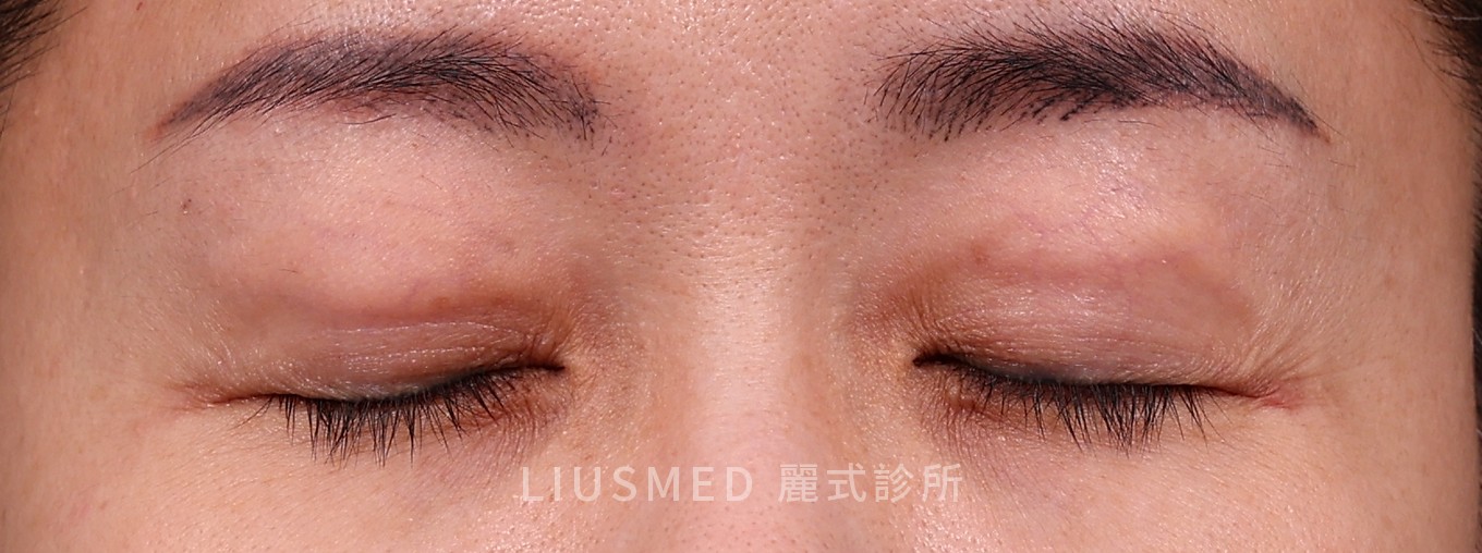
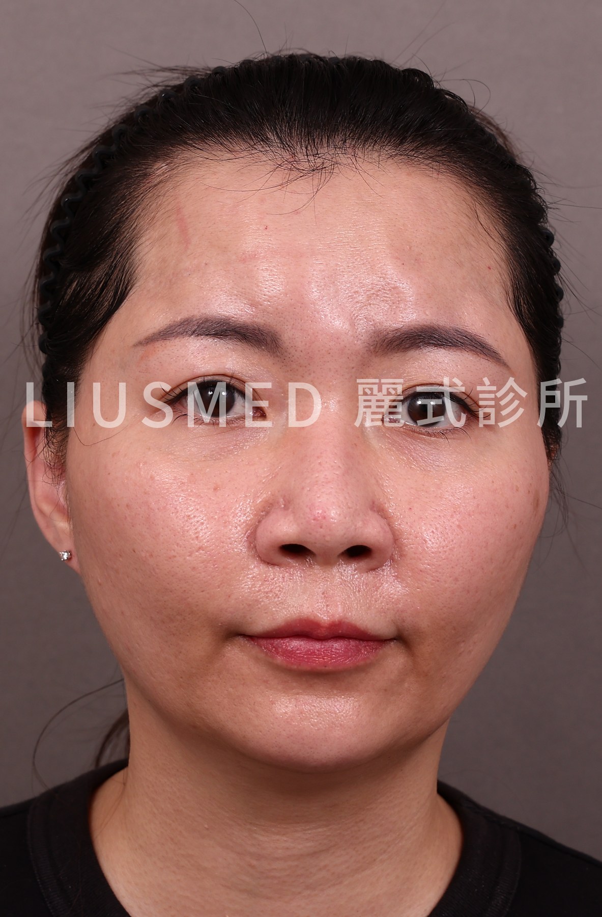
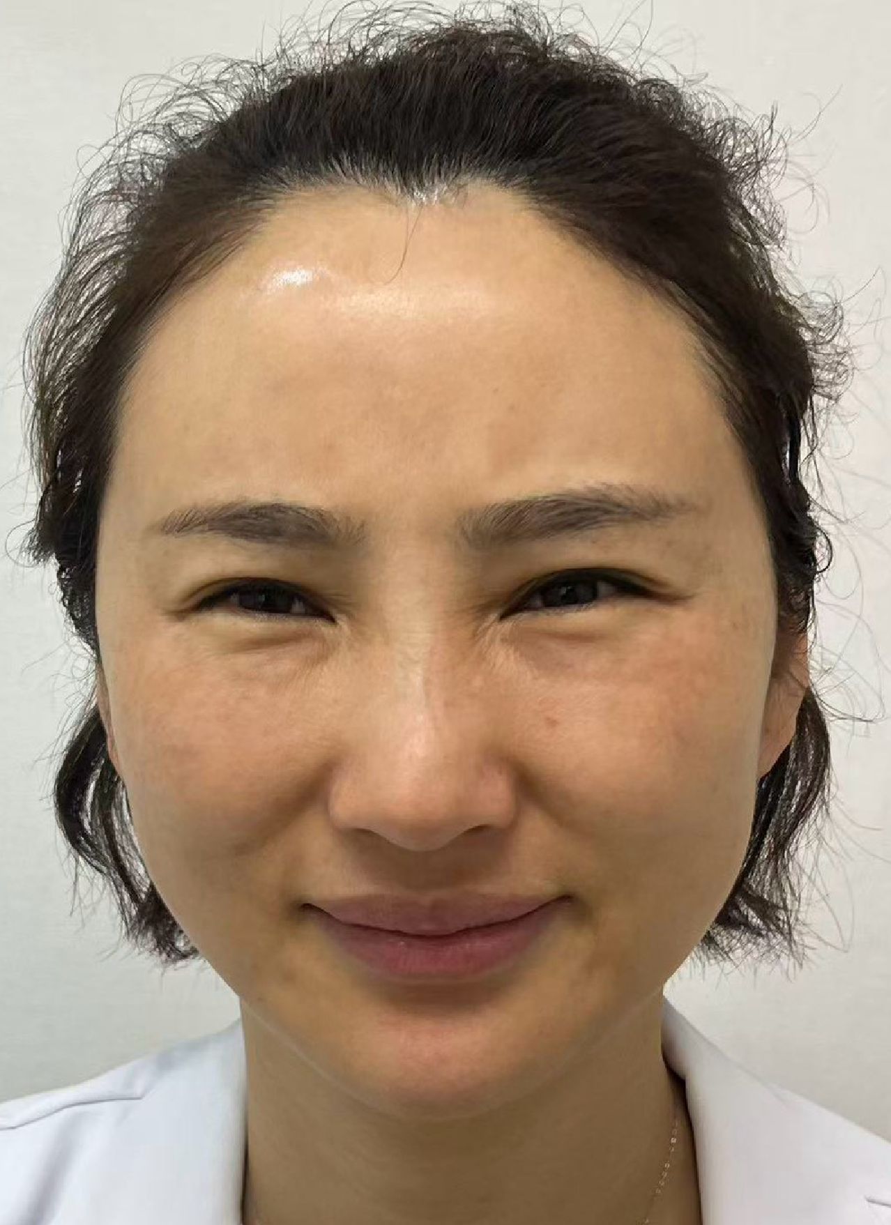
"There's a strip along my smile line, and it feels hard. They told me the dermis would blend in with my own tissue. Why is it still there two years later?"
I hear this more and more often in consultations. Some people had allograft dermis placed, others had a piece of their own dermis taken from elsewhere on the body. The most common site is the nasolabial fold, followed by the under-eye area and the aegyo-sal (the small roll under the lower lash line). Some had it on the nasal bridge, and some had it used to fill a depressed scar.
Let me be clear first: dermis is not a bad material. On the forehead I actually like injectable dermal powder, because it sits smoothly and rarely overgrows. What goes wrong is usually where it was placed, how thick it was and how many layers were stacked. Placed poorly, it turns into a whole strip or block you can feel and see.
This article first helps you work out which kind you have, then lays out what each of four options really costs.
Key insight: Allograft dermis, injectable dermal powder and autologous dermis are three different things. Work out which one you have first, or every decision after that can head in the wrong direction.
First, Sort It Out: Implanted Sheet, Injected Powder, or Your Own Dermis
These three names sound alike, and they are managed very differently.
← Swipe to see more →
| Allograft dermis (sheet, strip, block) | Injectable dermal powder | Autologous dermis | |
|---|---|---|---|
| What it is | Donor skin with the cells removed, leaving the dermal scaffold, cut into a sheet, strip or block | The same scaffold milled into particles and mixed with fluid for injection | A small piece of your own dermis, sometimes with a layer of fat attached (dermis-fat graft) |
| How it goes in | Placed under the skin as a whole piece through a small incision during surgery | Injected with a needle | Placed as a whole piece like allograft dermis, plus a second wound where it was harvested |
| What a problem looks like | A whole strip or block that sticks out, with an edge you can feel | Localized hardening or a clump | Much like allograft dermis |
| Related reading | This article | Dermal powder hardening and fat calcification | This article |
When patients say AlloDerm (acellular dermal matrix) or name another brand, some mean a sheet and some mean a milled, injectable version. The same name does not mean the same form went in. If the original records are gone, ultrasound can usually tell whether you are looking at the edge of a single sheet or scattered particles.
This article is about implanted allograft dermis and autologous dermis. If what you had was dermal powder, the approach is closer to that for ordinary filler lumps, and the article linked above is the better starting point.
Why It Turns Into a Ridge
After dermis goes in, the body absorbs part of it. So surgeons often place it a little thicker on purpose, leaving room for that loss. A systematic review of 10 studies and 324 rhinoplasty patients concluded that overcorrection should be considered during surgery to deal with partial resorption afterward (PMID 38345943).
The trouble is that resorption is neither exact nor even. In a long-term follow-up of 25 secondary rhinoplasty patients, 45% had partial resorption, more over the bony bridge and less over the tip (PMID 16327615). The segment that absorbs less stays behind and becomes a ridge.
What I see in clinic is usually a few of these stacked together:
- The graft was thicker than needed, and the expected resorption never happened.
- One segment absorbed a lot and the next absorbed little, so the surface became uneven.
- The graft shifted after it was placed. Autologous dermis does this too: in a study of 331 dermis-fat grafts used in revision rhinoplasty, 25 cases needed another revision, 9 of them for deviation (PMID 32997238).
- It was placed where the skin is very thin, such as under the eye, so the edges were always going to be hard to hide.
To put it plainly, the material is usually not the problem. The problem is how it was placed and whether it suited that spot.
Nasolabial Fold, Under the Eye, Nasal Bridge, Depressed Scars: What Each Looks Like
Nasolabial fold
This is the site I see most. Some patients originally had a nasal-base augmentation, a procedure sometimes marketed in Asia as "aristocrat surgery." The usual description: it looks fine at rest, but when they smile a firm strip rises above the fold, or the two sides sit at different heights.
Under the eye and aegyo-sal
The skin here is the thinnest. You get a horizontal bulge with a visible edge that stands out more when smiling. This area is often mixed up with under-eye filler problems; see revising an overfilled aegyo-sal.
Nasal bridge
Most often an extra ridge along the bridge, or the whole strip leaning to one side.
Depressed scars
This one is different. What used to be a dip now bulges, and the edge is more noticeable than the original scar.
The nasolabial fold has plenty of filler problems of its own. If you are not sure whether that strip is dermis or filler from an earlier injection, start with how to tell them apart in a hard strip along the nasolabial fold, then confirm with ultrasound.
Four Options and How to Choose
← Swipe to see more →
| Approach | What it can do | Cost or limits |
|---|---|---|
| Wait and see | Volume still falls over the first six months after placement | After that, what remains has usually stabilized; a ridge still there after a year rarely resolves on its own |
| Fill around it with hyaluronic acid | Raises the low areas so the height difference looks smaller | Adds another layer of volume; the problem is covered, not reduced |
| Open surgical removal | An encapsulated graft can come out whole, capsule included | Requires cutting the skin and leaves a scar; tightly fused grafts are hard to clear; the spot may sink once the strip is gone |
| Ultrasound-guided pinhole reduction | No skin incision; only the protruding part is removed | Not easy whether the graft is fused or not; may take more than one session; no promise of 100% |
Wait and see
In a study of AlloDerm sheets, volume decreased over the first 6 months and then stabilized over the following 6 months, and biopsies showed the body's own cells gradually moving into the graft (PMID 11368660). In another secondary rhinoplasty follow-up, the nasal contour did not change between the first and second year (PMID 16327615).
So if the graft went in recently, watching it for a while can be reasonable. If it has been a year or two and the strip is still there, waiting longer is unlikely to help.
Fill around it with hyaluronic acid
This is the fastest way to make the height difference look smaller. The logic, though, is to add more: the ridge stays, and another layer goes on beside it. The total volume in the face keeps going up, and it easily ends with the whole area looking puffy.
Open surgical removal
Surgery is not without reason. Most dermal grafts develop a capsule, and an encapsulated graft is easier to take out whole in surgery. But it requires an incision in the skin, and on the face, that scar stays on the face.
A minority of grafts also become tightly fused with the surrounding tissue, and those are hard to clear even with surgery. Once the whole strip is out, the area it was holding up is suddenly empty, and a new dent often appears. I go through what excision costs on the face in more detail in scarring and dents after surgical excision.
Ultrasound-guided pinhole reduction
This is how I do it. First I use ultrasound to see which layer the graft is in, how thick it is and where its edges are. Then, through a pinhole of about 2 mm, I remove only the part that protrudes.
What sets this apart from surgery is that the thickness removed can be controlled. If a strip was placed too thick, for example, a first session usually takes off about 30 to 50 percent, and the surface becomes much smoother.
I also have to be honest: whether the graft is tightly fused or not, working on dermis through a pinhole is not easy. It is not guaranteed to be done in one go, it sometimes takes more than one session, and I cannot promise 100%. For how ultrasound-guided single-pinhole removal works in general, see how ultrasound-guided pinhole extraction is done.
Key insight: Surgery can take an encapsulated graft out as a whole strip, at the cost of an incision and scar on the face, plus the dent left once it is empty. Pinhole reduction does not cut the skin and can deal with only the protruding part, but it takes time and patience and may be done over several sessions.
Why I Don't Take the Whole Strip Out
Taking material out and putting it in follow the same logic.
When we inject, we want overall harmony and avoid putting too much in one spot, which is how overfilling happens. Taking it out is no different. After overfilling, you do not need to remove 100% of what is there. You follow the natural line and shape the part that remains until it is smooth and even.
Dermis shows this better than anything. The surgical approach is to open it up and take the whole strip out, and the spot it came from easily ends up dented. We remove only what protrudes, so that the line comes back level with its surroundings. When the proportions are right and the lines are natural, the face looks good.
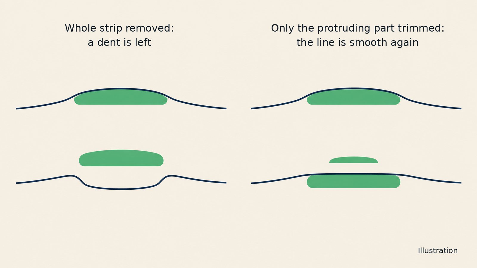
So when I assess a patient, I look at the lines of the whole face: where there is extra, and how much has to come off for it to sit level with the rest.
When Not to Rush
- It is red, swollen, warm, painful, or draining pus: treat the infection first. There are reports of allograft dermis in the nasal tip becoming infected and having to be removed surgically (PMID 26894006). Reduction is not appropriate until the infection is under control.
- It went in less than six months ago: the volume is still changing. Watch it first, and assess once it has stabilized whether to treat it and how much.
- You are not actually sure what is in there: what was placed for a nasal-base augmentation or a nose job is not necessarily dermis. It may be a silicone implant, cartilage or filler injected earlier. These are handled completely differently, so confirm with ultrasound first.
What Ultrasound Looks For First
Dermal grafts are visible on ultrasound. A Korean study of folded dermal grafts in rhinoplasty used ultrasound to follow the grafts and found a 38% loss of height within 6 months (PMID 40728913).
When I assess, I look at a few things: which layer it is in, how thick it is, whether its edges are clear, whether there is a capsule, whether there is fluid or inflammation around it, and how close it lies to important vessels and nerves. Only once those are clear can I tell whether it can be reduced, how much, and whether it should be split into sessions.
Recovery and Realistic Expectations
The pinhole is small, but this is still a removal, and there will be swelling and bruising. We do not claim the recovery is short.
- Swelling and bruising fade over about 2 to 3 weeks.
- The pinhole heals in about 2 weeks and is usually hard to notice after that, though this still depends on how your skin heals.
- The overall result takes about 3 months to settle, and this varies from person to person.
Complete removal is not guaranteed. The aim is to improve as much as can be done safely and restore a natural contour. If more than one session is needed, they are usually about 3 months apart.
Frequently Asked Questions
Will allograft dermis absorb on its own?
Part of it, but not all of it. In a study of AlloDerm sheets, volume decreased over the first 6 months after placement and then stabilized (PMID 11368660). A strip still there after a year usually will not go away by itself.
Can allograft dermis be dissolved with hyaluronidase?
No. Hyaluronidase breaks down hyaluronic acid, and dermis is mostly a collagen scaffold, so the enzyme does nothing to it. Repeated injections only keep irritating the tissue.
Autologous dermis is my own tissue. Can it still bulge, and can it be removed?
Yes. Autologous dermis can also absorb unevenly, shift or be placed too thick. It is handled much the same way as allograft dermis, and it can likewise be located with ultrasound and reduced through a pinhole.
Does all of it have to come out?
No. The goal is smooth lines and balanced proportions, not taking every bit of dermis out. Often, removing just the protruding portion improves the appearance a great deal, and taking out too much makes a dent more likely.
Isn't surgery more thorough?
An encapsulated graft can indeed come out whole with surgery, but that means an incision, a scar, and possibly a dent where it used to be. A minority that are tightly fused are hard to clear even with surgery. Whether surgery makes sense depends on what matters to you and what the graft looks like on ultrasound.
Will removal leave a scar?
The pinhole is about 2 mm and needs no stitches. It heals in about 2 weeks and fades over about 2 months, and in most people it becomes very hard to see, though scarring still depends on your own healing.
Can I have filler or a graft again afterward?
It can be discussed, but there is no hurry. We usually wait about 3 months for the tissue to settle, then review the overall lines at a follow-up visit before deciding whether anything is needed.
Want to Check Your Own Situation?
If you have had allograft or autologous dermis placed and can now feel a strip or see a block, the first step is simple: find a doctor who uses ultrasound to look at implants under the skin, and confirm what it is, which layer it is in and how thick it is.
I have spent many years removing and repairing fillers, and I have taken on many patients whose earlier treatment elsewhere did not turn out well. To talk through your situation, start with booking a consultation, or read the overview of filler repair first.
References
- Tang Y, Han X, Li J, et al. A Systematic Review of Technique, Satisfaction, and Complications of Acellular Dermal Matrix in Rhinoplasty. J Craniofac Surg. 2024. doi:10.1097/SCS.0000000000010000. PMID: 38345943.
- Gryskiewicz JM. Waste not, want not: the use of AlloDerm in secondary rhinoplasty. Plast Reconstr Surg. 2005;116(7):1999-2004. PMID: 16327615.
- Kim JH, Ko HS, Park SW. Using Dermofat Grafting in Revision Rhinoplasty. Aesthetic Plast Surg. 2021;45(2):617-625. PMID: 32997238.
- Sclafani AP, Romo T 3rd, Jacono AA, et al. Evaluation of acellular dermal graft (AlloDerm) sheet for soft tissue augmentation: a 1-year follow-up of clinical observations and histological findings. Arch Facial Plast Surg. 2001;3(2):101-103. PMID: 11368660.
- Lee KH. Infection in the Nasal Tip Caused by Acellular Dermal Matrix. Plast Reconstr Surg Glob Open. 2015;3(12):e581. PMID: 26894006.
- Kim DG, Kim KH, Cha HG, et al. Volume Retention of Folded Dermal Graft After Augmentation Rhinoplasty. J Craniofac Surg. 2025. doi:10.1097/SCS.0000000000011723. PMID: 40728913.
Editorial Review: This article was written by Dr. Ta-Ju Liu based on clinical experience and the published literature. It is for patient education and does not replace an in-person consultation. The open surgical removal cited here is the approach described in the literature; our clinic treats these cases with ultrasound guidance through a single pinhole. Whether this is suitable for you, and how it would be done, is decided only after an in-person consultation and ultrasound assessment.