"Why Not Just Cut It Out?"
"My doctor says dissolving and steroids have failed, so the primary option left is to cut it out." At FILLER REVISION, we hear this from patients who have been told surgical excision is their last resort — often by practitioners unaware that minimally invasive, ultrasound-guided alternatives exist. Many of these patients arrive expecting to need open surgery, only to learn that their lump can be removed through a single pinhole without visible scarring.
Traditional surgical excision is a method that works but at a high cost — it can indeed remove the lump, but the accompanying scar, tissue deficit, and recovery period often leave patients trading one problem for another of equal or greater severity.
How Is Traditional Excision Surgery Performed?
Surgical Steps
- A skin incision is made over or near the lump (typically 1-3 cm)
- Tissue is separated layer by layer until the lump is reached
- The lump and surrounding tissue are excised together
- The wound is closed with layered sutures
- Post-operative suture removal and wound care
Why Is Open Surgery on the Face Particularly Risky?
← Swipe to see more →
| Risk Factor | Description | Severity |
|---|---|---|
| Visible scarring | Any incision on facial skin leaves a permanent scar | High |
| Nerve damage | Incision may sever branches of facial nerves | High |
| Tissue deficit | Excision extent may exceed what is necessary, leaving depressions | Medium-high |
| Asymmetry | Unilateral excision creates side-to-side imbalance | Medium |
| Extended recovery | Facial open surgery requires 2-4+ weeks recovery | Medium |
| Infection risk | Open surgical wounds carry higher infection risk | Medium |
Scarring: The Cruelest Cost of Facial Surgery
Why Are Facial Scars Particularly Conspicuous?
The face is the most scrutinized area in human interaction. Even with the most refined suturing technique, facial incisions will leave some degree of scarring. Scar severity depends on multiple factors:
- Incision placement: Incisions that do not follow relaxed skin tension lines produce more visible scars
- Incision length: The incision needed for lump removal is typically 1-3 cm
- Individual constitution: Patients prone to hypertrophic scarring or keloid formation face worse outcomes
- Post-operative care: Infection or excessive tension worsens scarring
Key Insight: At FILLER REVISION, we remind patients of this principle: the original concern was "I have an unsightly lump on my face." If the solution leaves a permanent scar, for many patients this is not a true resolution. The goal of treating filler complications should be to restore natural appearance as much as possible — not to replace one problem with another.
The Trap of Over-Excision
In traditional surgery, surgeons habitually ensure "clean margins," often excising a safety border around the lump. In tumor surgery, this is reasonable and necessary. But for filler lumps, this mindset can cause unnecessary tissue sacrifice.
Filler lumps are not tumors—they do not spread or metastasize. Over-excision only creates a larger tissue deficit, and on the face, this deficit directly manifests as depression and asymmetry.
For more analysis on why encapsulated filler cannot rely on dissolution, see: Encapsulation: Why Dissolvers Fail.
The FILLER REVISION Approach: When Standard Treatment Points to Surgery
At FILLER REVISION, we see patients every week who were told their only remaining option is surgical excision. In the vast majority of these cases, our ultrasound evaluation reveals that minimally invasive extraction is not only feasible but preferable. The key difference is imaging: traditional surgery relies on direct visualization through an incision, but ultrasound provides equal or better visualization through the skin — without cutting. Our pinhole extraction technique removes filler and capsule tissue under real-time ultrasound guidance, achieving thorough removal without the scarring, tissue deficit, and prolonged recovery of open surgery. For the small number of cases that genuinely require open surgery, our pre-procedure ultrasound still provides essential information for planning the smallest possible incision and minimizing tissue sacrifice.
Ultrasound (Ultrasonography)-Guided Minimally Invasive Extraction vs. Traditional Open Surgery
← Swipe to see more →
| Comparison | Traditional Open Surgery | Ultrasound-Guided Extraction |
|---|---|---|
| Entry size | 1-3 cm incision | Single pinhole (1-2mm) |
| Scarring | Permanent visible scar | Nearly invisible |
| Visual guidance | Direct visualization | Real-time ultrasound |
| Surrounding tissue damage | Layer-by-layer dissection required | Precise path preserving normal tissue |
| Recovery time | 2-4 weeks | Several days |
| Anesthesia | May require general or regional | Local anesthesia |
| Material identification | Confirmed only during surgery | Identified before procedure |
| Residual risk | Eyes may miss deep material | Ultrasound confirms clearance |
Key Insight: The reason ultrasound-guided minimally invasive extraction can achieve more thorough filler removal while preserving more normal tissue lies in its ability to see. Ultrasound provides not a blurry outline but precise information about tissue layers, material characteristics, and real-time instrument position.
When Might Traditional Surgery Still Be Necessary?
Objectively, minimally invasive extraction is not appropriate for every situation. In a very small number of scenarios, traditional surgical excision may still be necessary:
- Extremely extensive filler spanning multiple tissue planes: Some historical large-volume injections (such as early unregulated injectable procedures) involve material that has spread extensively across multiple tissue layers
- Severe infection requiring open drainage: Deep abscesses require adequate drainage channels
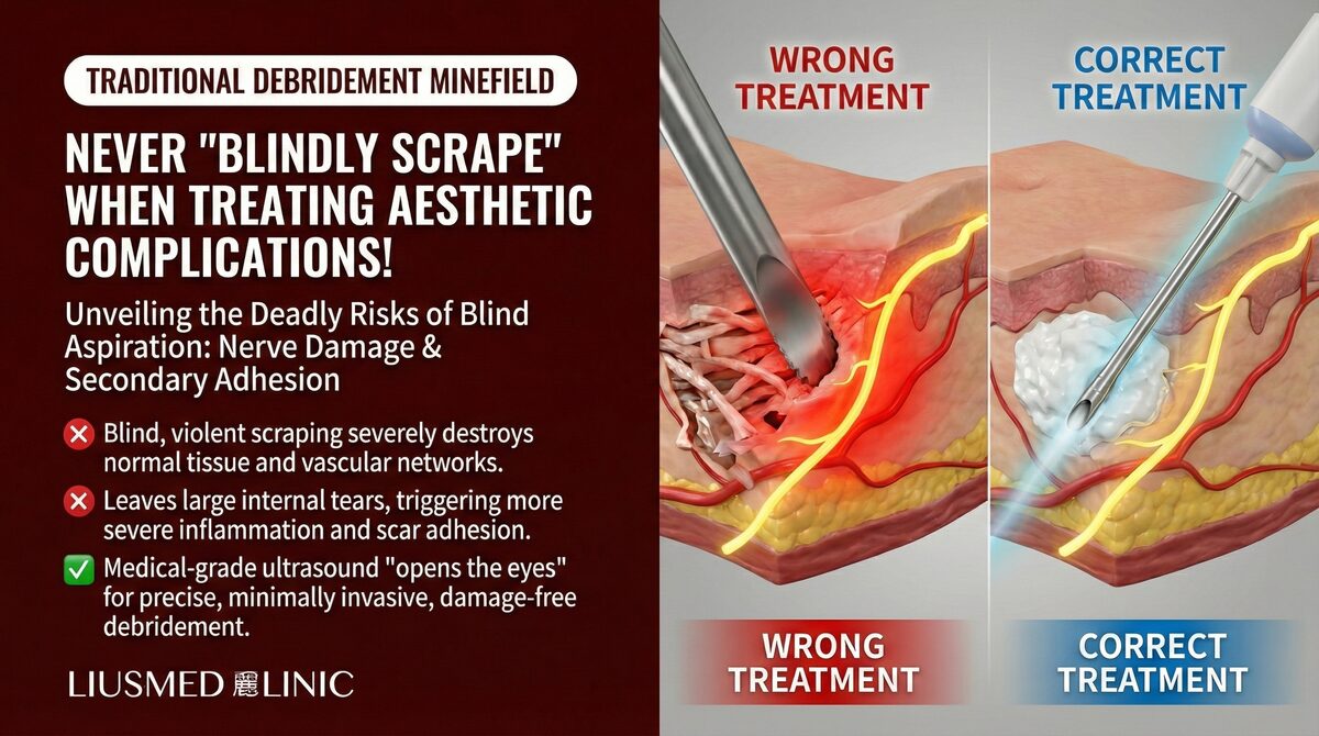
- Associated tissue necrosis requiring debridement: Areas of established tissue necrosis require thorough debridement
However, even in these situations, ultrasound evaluation remains an indispensable first step—to determine the extent of the problem and formulate a surgical plan.
Core Advantages of Minimally Invasive Technique
The Significance of a Single Pinhole
"One pinhole" is not merely marketing language—it represents a fundamental shift in treatment philosophy:
- Minimized invasion: The entry point is only a pinhole, reducing surface skin damage to the absolute minimum
- Internal precision: Under real-time ultrasound guidance, precise filler extraction is performed through this pinhole
- Tissue integrity preserved: No layer-by-layer dissection and suturing required; surrounding tissue structure is maintained
- Rapid recovery: No swelling and healing burden from a large incision
The Critical Role of Pre-Procedure Ultrasound
Before every extraction procedure, a complete ultrasound scan is performed. This is not merely a "quick look" but the creation of a detailed treatment map:
- Precise filler location, depth, and extent
- Material characteristics and degree of encapsulation
- Pathways of surrounding critical vessels and nerves
- Optimal entry path planning
For more detailed information on the extraction technique, see: Filler Lump Extraction Technique Explained.
Recommendations Before Making a Decision
If you are considering surgical treatment for facial filler lumps, we recommend the following before making a final decision:
- Get an ultrasound evaluation first: Understand the exact material, location, and extent of the lump
- Assess minimally invasive feasibility: The vast majority of cases can be managed with minimally invasive approaches
- Understand scarring risks: If traditional surgery is ultimately needed, fully understand the scar location and severity
- Seek a second opinion: Especially when the first practitioner recommends direct surgical excision
We recommend starting with a comprehensive ultrasound evaluation. Schedule a consultation and let us find the most appropriate treatment plan for your specific situation.
Conclusion
If you have been told surgical excision is your primary option, FILLER REVISION offers a second opinion and, in most cases, a scar-free alternative. Our ultrasound-guided pinhole extraction achieves thorough filler removal without the incision, scarring, and tissue sacrifice of traditional open surgery.
Before agreeing to any procedure that will leave a permanent scar on your face, explore all available options. Book a consultation →
Frequently Asked Questions
My doctor says dissolving and steroids failed, so the only option left is to cut the lump out. Is surgery really my last resort?
Not necessarily. At FILLER REVISION, many patients who were told surgical excision was their only remaining option turn out, after an ultrasound evaluation, to be candidates for minimally invasive pinhole extraction instead — which is feasible in the vast majority of cases. Before agreeing to open surgery, the article recommends getting an ultrasound evaluation first and seeking a second opinion, especially when the first practitioner recommends direct surgical excision.
How big is the incision for surgery, and will the scar be permanent?
Traditional surgical excision of a filler lump typically requires a 1-3 cm facial incision, and any incision on facial skin leaves a permanent scar. By contrast, ultrasound-guided pinhole extraction uses a single entry point of only 1-2mm, with nearly invisible scarring. The article notes facial scars are especially conspicuous because the face is the most scrutinized area in human interaction.
The surgeon wants to cut out extra tissue around the lump for 'clean margins.' Is that necessary for filler?
Filler lumps are not tumors — they do not spread or metastasize, so the 'clean margins' approach used in tumor surgery is not required. Over-excising a safety border only creates a larger tissue deficit, which on the face directly shows up as depression and asymmetry. The article describes this habit of over-excision as an unnecessary tissue sacrifice for filler lumps.
How long is recovery, and what kind of anesthesia is used compared with open surgery?
Traditional facial open surgery generally requires 2-4 weeks of recovery and may call for general or regional anesthesia. Ultrasound-guided pinhole extraction is done under local anesthesia, with recovery measured in several days rather than weeks because there is no large incision causing swelling and a heavy healing burden. The exact plan for your situation is discussed during a LINE or in-person consultation.
Are there any situations where traditional open surgery is still genuinely needed?
Yes, in a very small number of scenarios. The article lists extremely extensive filler spanning multiple tissue planes (such as early unregulated large-volume injections), severe infection requiring open drainage of a deep abscess, and associated tissue necrosis requiring debridement. Even in these cases, ultrasound evaluation remains an indispensable first step to determine the extent of the problem and plan the smallest possible incision.
Why is ultrasound considered as good as or better than seeing the lump directly during surgery?
Traditional surgery relies on direct visualization through an incision, but ultrasound provides equal or better visualization through the skin without cutting. It shows precise information about tissue layers, material characteristics, and the real-time position of instruments — identifying the material type and extent before the procedure even begins. This is what lets thorough removal happen while preserving more normal tissue.