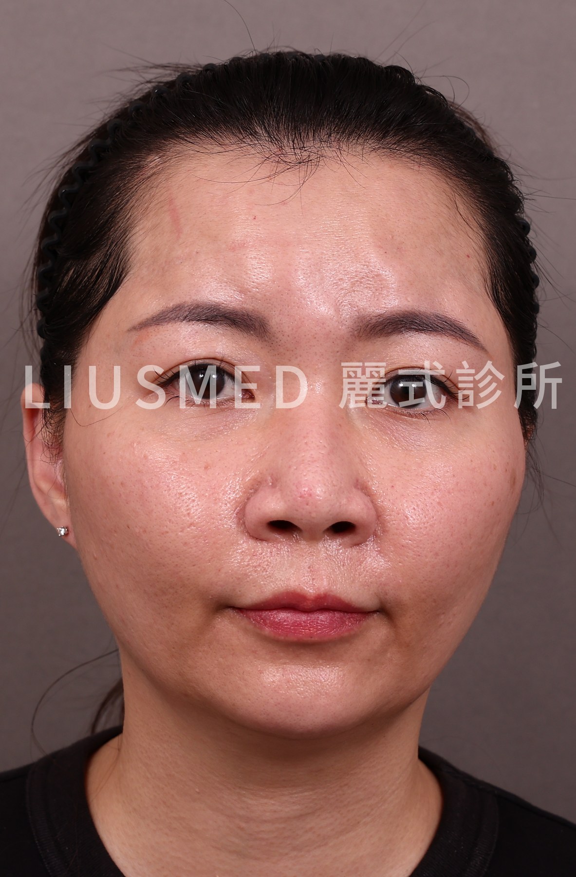
Injecting collagen stimulators as a "skin booster" — shallow, full-face, microdroplets packed close together — has become one of the most popular skin-quality treatments of the past two or three years. In clinic, more and more people come not because "a bolus of filler turned hard," but because "a whole patch of my cheek or forehead feels grainy, like little beads under the skin." This is one of the most overlooked, and hardest to dissolve, forms of collagen stimulator lumps.
This article is not about the decision of "should I get a skin booster at all" (a separate topic, linked at the end). It focuses on what happens after the nodules have already formed: what they actually are, why hyaluronidase can't dissolve them, what they look like on ultrasound, and why physical extraction is so often the practical option.
Skin booster lumps not going away: what's normal, and when to get them checked
It depends on when the bumps appeared and what was injected. A rough guide from the literature:
- The first few days. Small bumps, swelling and bruising right after a skin booster are expected; for HA boosters such as Profhilo, a systematic review found they usually settled within 72 hours.
- Still there after about two weeks. Problems lasting longer than two weeks are often linked to technique, for example a long-lasting product placed too superficially.
- Appearing a few weeks later. With PLLA, small papules and nodules tend to show up several weeks after injection; early implant nodules usually appear 2 to 4 weeks after.
- Appearing months or years later, at all the injected spots at once. That pattern points toward a granuloma or delayed reaction, which can appear months to years after injection.
- Red, warm, tender or growing. Get this seen promptly, whatever the timing.
Will they go away on their own? That depends on the type. In the PLLA literature, small papules you can feel but not see, and that don't hurt, often settle by themselves. Firmer nodules are different: in 55 patients with collagen-stimulator complications reported by ultrasound specialists, only five cleared completely despite saline, hyaluronidase, diluted steroid and energy-based treatments. The two groups can't be compared directly (one is everyday patients, the other cases reported by ultrasound specialists), but side by side they show why "just wait" works for some lumps and not others. If yours are past the two-week mark, or keep coming back, an ultrasound assessment is the practical next step to find out which kind you have.
Which skin booster was it? Why that decides whether the lump can dissolve
"Skin booster" describes how a product is injected, shallow and spread over a wide area, not what is in it. The material decides whether hyaluronidase has anything to act on:
← Swipe to see more →
| What was injected | Can hyaluronidase dissolve it? | Read more |
|---|---|---|
| Pure hyaluronic acid boosters (including Profhilo-type products) | Yes | Why hyaluronidase sometimes fails |
| HA mixed with a collagen stimulator (e.g. Juvelook, HArmonyCa) | Only the HA part | Juvelook / Lenisna nodules, HArmonyCa nodules |
| Human-derived dermal matrix (RE2O / phADM) | Only any HA added when it was mixed; the matrix particles don't dissolve | What RE2O is and its complications, RE2O bumps and nodules |
| Polynucleotides (PN / PDRN) | Too little data to say | — |
| Diluted collagen stimulators (Sculptra, AestheFill, Ellansé, Radiesse) | No | AestheFill nodule treatment ladder, Sculptra lumps after steroids, Ellansé delayed nodules |
In the 55 patients above, hyaluronidase only helped when HA was part of the product. For PN boosters the evidence is thin: a systematic review found nine studies of low to moderate quality covering 219 patients, so we say "too little data" rather than guess. Placing a collagen stimulator shallow and over a wide area is a real technique (hyperdilute CaHA is placed this way on purpose), which is why these lumps now turn up after treatments sold as skin boosters.
1. What is actually injected in a "collagen stimulator skin booster"?
To understand why it turns into lumps, you first have to separate two ideas: collagen stimulators work in a completely different way from hyaluronic acid.
Hyaluronic acid (HA) is "fill" — it is the volume; you inject it and it sits there holding space. Collagen stimulators are "induce" — the product itself is just a carrier and particles; the real volume comes from the collagen your own body grows in response to it. The common base materials:
- PLLA (poly-L-lactic acid; the active ingredient in Sculptra): gradually stimulates deep collagen growth, originally designed for full-face contour support.
- PCL (polycaprolactone; the active ingredient in Ellansé): a dual mechanism of immediate fill plus long-lasting collagen stimulation, with stronger support.
- PDLLA (poly-D,L-lactic acid; the active ingredient in AestheFill): softer texture, uniform particles, marketed for skin-quality improvement.
- CaHA (calcium hydroxylapatite; the active ingredient in Radiesse): mineral-based particles giving immediate three-dimensional effect.
The crux of the problem is not the material itself but that the way it is injected has changed. These products were mostly designed for deep, point-specific, appropriately diluted injection. The recent "skin-booster trend" — including hybrid products like Juvelook that combine hyaluronic acid with collagen-stimulating components — has reshaped them into shallow, wide-area, high-density microdroplets. The same material, placed at the depth it was meant for, gives skin quality and support; placed too shallow and too dense in the dermis, it can become a row of beads you can feel.
Key point: Whether a collagen stimulator becomes a nodule is usually less about "good or bad material" and more about "depth and distribution." Injected as shallow, wide-area microdroplets, the collagen-stimulating particles stay in the superficial dermis, and the body encases each droplet in collagen and fibrous tissue — producing multiple, scattered small lumps.
How common is this? In a meta-analysis of biostimulators (CaHA, PLLA and PCL) used in the usual way, nodules developed in about 5% of patients (95% CI 2–10%); there is no separate figure for the skin-booster technique. The causes the PLLA literature lists are familiar: incorrect reconstitution or dilution, uneven distribution, injecting too superficially and skipping post-treatment massage. Not every study puts technique first, though: a Brazilian series of 55 cases concluded the complications related more to the products' properties than to technique. For PLLA, true inflammatory granulomas are much rarer, around 0.01–0.1%.
2. How is this lump different from an HA lump?
This is the most important section, because it directly determines whether the lump can be dissolved.
An HA lump is mostly HA gel, so hyaluronidase (the enzyme that dissolves HA; to be used only after physician evaluation) can break it down. But a collagen-stimulator nodule is mostly your own collagen fibres encasing residual particles, sometimes with an accompanying granuloma (a chronic immune nodule the body forms around foreign material). Hyaluronidase cleaves the bonds of hyaluronic acid; against PLLA, PCL, PDLLA or CaHA particles and the collagen fibres around them, it has nothing to act on at all.
In other words, injecting hyaluronidase into a collagen-stimulator lump isn't "too low a dose" — it's "aimed at the wrong target."
← Swipe to see more →
| Comparison | HA lump | Collagen stimulator lump |
|---|---|---|
| Main composition | HA gel | Own collagen fibres + residual particles (± granuloma) |
| Typical feel | Elastic, well-defined mass | With microdroplet technique, often multiple, scattered, grainy small nodules |
| Time to appear | Relatively soon after injection | Often weeks to months later, becoming clearer as collagen grows |
| Response to hyaluronidase | Dissolves | No target — does not dissolve |
| Main treatment path | Hyaluronidase first, extraction if needed | Physical extraction after ultrasound identification |
Key point: "Hyaluronidase had no effect" is itself an important clue — it often means the material is not HA but a collagen stimulator or a mixed product. Continuing to add more hyaluronidase does nothing for the nodule, while repeatedly irritating the surrounding tissue.
Why collagen stimulators trigger this immune and fibrotic response is a deeper topic in its own right; for the detail, see our explanation of collagen stimulator mechanisms and risks. Here we stay focused on "what to do once the nodule has already formed."
Red, warm and tender, or painless and hard? Two different kinds of nodule
Non-inflammatory nodules usually come from the product building up or spreading unevenly, or from being placed at the wrong depth; they feel firm but don't hurt. Inflammatory nodules come with warmth, redness, tenderness and swelling. One review of PLLA nodules sorts them into four types: non-inflammatory, inflammatory-immune, infection or biofilm, and unusual presentations. The type decides the order of treatment. If there is infection or active inflammation, that has to be brought under control first; removal comes after it, not instead of it. For swelling that settles and then flares again, see biofilm and recurring filler swelling.
3. What do collagen stimulator nodules look like on ultrasound?
"You can only treat safely what you can see" is a consistent principle at FILLER REVISION. Under ultrasound guidance, different materials show distinct imaging features, and these guide both identification and localisation:
- HA: mostly a hypoechoic-to-anechoic cystic structure with relatively clear borders.
- PLLA / PDLLA microdroplets: in the superficial dermis, often punctate, scattered hyperechoic signals — matching the microdroplet distribution: not one mass, but a sheet.
- CaHA: because of its mineral particles, shows a strong echo with posterior acoustic shadowing, a relatively recognisable feature.
- PCL: microsphere structures with echo changes from surrounding fibrosis.
A 2026 series of 62 patients described these contrasts in more detail: HA nodules shift from anechoic "jelly" to hypoechoic "stormcloud" patterns, PDLLA nodules appear as well-defined hyperechoic "snow globes", and PCL ranges from a fibrotic "dragon-beard candy" look to layered "water ripple" patterns. Separately, a small series of PCL granulomas described them as hypoechoic areas with bright surface dots and comet-tail artefacts.
The ultrasound signature of skin-booster microdroplet technique is often superficial, multiple and widely distributed — very different from the single mass left by traditional deep point injection, and this directly shapes the extraction strategy. The more widely scattered the small nodules, the more they call for precise localisation and one-by-one handling, rather than blind aspiration or blind scraping.
4. Why is physical extraction often the main option? The limits of steroids and 5-FU
For collagen-stimulator nodules, the common drug options are intralesional steroid (triamcinolone) or 5-FU (5-fluorouracil, an antimetabolite). Each has a role, and each has clear limits:
- Steroids: can suppress surrounding inflammation and temporarily soften and shrink the nodule, but do not remove the particles themselves. Repeated injection can also cause local skin and subcutaneous atrophy and depression — leaving another problem behind.
- 5-FU: has some inhibitory effect on collagen overgrowth and is often combined with steroid, but has limited effect on an already-formed particle-plus-fibrosis nodule and usually requires multiple sessions. For a fuller discussion, see the limits of 5-FU for collagen stimulators.
The literature points the same way. Lemperle's treatment review notes that steroids can be combined with antimitotic drugs such as 5-FU. A systematic review of foreign-body granulomas from collagen stimulators found intralesional steroid was the most used treatment (21.37%), yet symptoms were often resolved through surgical removal. A PDLLA management guideline states that these nodules do not respond to straightforward enzymatic treatment. Saline injection has occasionally helped: in one multicentre series, a single PLLA nodule resolved after two saline injections. If you've already had several rounds of 5-FU without result, see collagen stimulator lumps after 5-FU fails; for thinning after repeated injections, see skin atrophy after lump injections.
This is why, once a nodule has formed and drugs repeatedly fail, physical extraction becomes the more practical direction: precise localisation under ultrasound guidance, then removing the particles and the fibrous tissue encasing them directly — through the single-pinhole physical extraction approach our clinic has developed over many years — rather than relying on chemical dissolving or repeatedly suppressing inflammation. Whether and how something can be removed depends on the material type, the nodule depth and its distribution; for the removability of PCL products specifically, see can Ellansé be removed. If your nodules are the delayed-onset kind that PCL products such as Ellansé can leave behind, the full Ellansé nodule treatment ladder sets out the order of the options in detail.
Microdroplet, shallow, multiple nodules do demand more patience to extract: they are widely distributed and individually small, and the precision of localisation directly determines how completely they are removed and how well normal tissue is protected. That is exactly the value of identifying first with ultrasound and extracting second — you can read more in the filler repair service overview, or browse the collagen stimulator lump hub.
5. Reconstruction and follow-up after removal
Removal is not the end; what follows matters just as much.
On recovery, we don't claim it is short. The pinholes are small, but this is still removal, so swelling and bruising will happen: they usually settle in about 2–3 weeks, the pinholes heal in about 2 weeks, and the final result takes about 3 months, varying from person to person. Widely scattered nodules may be handled in zones or over more than one session, still through pinholes, and the aim is as much improvement as can be achieved safely, not a 100% clearance. How it works step by step is in ultrasound-guided pinhole extraction explained.
After collagen-stimulator microdroplet nodules are removed, the previously encased area is left with space, and there may be varying degrees of surrounding fibrosis. We do not rush to re-fill the same spot — letting the tissue recover, allowing inflammation to settle, and then confirming with follow-up ultrasound that no residual particles remain is the steadier sequence. Once the tissue is stable, we discuss — case by case — whether and how to rebuild volume and skin quality.
If you are still hesitating over "I already have collagen stimulator lumps, can I still get a skin booster or skin-quality treatment," that belongs to the pre-treatment decision; our sister clinic has a dedicated article on it: can you get a skin booster with existing filler lumps. This article stays focused on "how to identify and safely remove a nodule that has already formed."
Key point: In handling collagen-stimulator nodules, sequence matters more than speed — see clearly first (ultrasound identification), then decide the method (physical extraction primarily, drugs as support), and only then discuss reconstruction. Skipping identification and going straight to blind injection or blind aspiration is usually where things start to get complicated.
Injecting collagen stimulators "as a skin booster" is a trend of recent years, and it has made these shallow, multiple nodules a common concern in clinic. The frustration is real — hyaluronidase won't dissolve them, drugs only suppress, and the surface looks like a whole patch of grain. But with correct identification and precise localisation, in most cases they can be handled safely.
If you are troubled by nodules after collagen-stimulator microdroplet injection, you are welcome to use our online evaluation and consultation booking, where Dr. Ta-Ju Liu can assess your actual situation with ultrasound and discuss the most suitable approach with you.
Frequently Asked Questions
I had hyaluronidase injected but the lumps didn't go away — why?
Hyaluronidase only breaks down hyaluronic acid. A collagen-stimulator nodule is mostly your own collagen fibres encasing residual PLLA/PCL/PDLLA/CaHA particles, so the enzyme has nothing to act on — it is not a matter of too low a dose, but of aiming at the wrong target. In fact, the fact that hyaluronidase had no effect is itself an important clue that the material may not be HA. Continuing to add more hyaluronidase does nothing for the nodule while repeatedly irritating the surrounding tissue.
How can you tell whether my lumps are hyaluronic acid or a collagen stimulator?
Ultrasound is the key. On ultrasound, HA usually appears as a hypoechoic-to-anechoic cystic structure with relatively clear borders, while collagen-stimulator microdroplets (PLLA/PDLLA) in the superficial dermis show punctate, scattered hyperechoic signals, and CaHA shows a strong echo with posterior acoustic shadowing. The clinical history also helps: if hyaluronidase had no effect, that itself often points to a collagen stimulator or mixed product rather than HA.
Can steroid or 5-FU injections clear the nodules instead of removing them?
These drugs have a role, but clear limits. Steroids can suppress surrounding inflammation and temporarily soften and shrink a nodule, but they do not remove the particles themselves, and repeated injection can cause local skin and subcutaneous atrophy and depression. 5-FU has some inhibitory effect on collagen overgrowth and is often combined with steroid, but has limited effect on an already-formed particle-plus-fibrosis nodule and usually needs multiple sessions. Once a nodule has formed and drugs repeatedly fail, physical extraction that addresses the root becomes the more practical direction.
Why are my lumps lots of small scattered beads instead of one hard mass?
It usually comes down to depth and distribution rather than whether the material was good or bad. When a collagen stimulator is injected as a skin booster — shallow, wide-area, high-density microdroplets — the particles stay in the superficial dermis, and your body encases each droplet in collagen and fibrous tissue, producing multiple scattered small lumps. This differs from the single mass left by traditional deep point injection, which is why on ultrasound the pattern is typically superficial, multiple and widely distributed.
After the nodules are removed, can I get a skin booster or re-fill the same spot right away?
We do not rush to re-fill the same spot. After removal the previously encased area is left with space and there may be surrounding fibrosis, so the steadier sequence is to let the tissue recover, allow inflammation to settle, and confirm on follow-up ultrasound that no residual particles remain. Only once the tissue is stable do we discuss, case by case, whether and how to rebuild volume and skin quality. Whether you can have a skin booster while you still have existing lumps is a separate pre-treatment decision covered in a dedicated sister-clinic article.
How long do bumps after a skin booster last, and when should I get them checked?
Small bumps, swelling and bruising in the first few days are expected and usually settle quickly. Bumps still there after about two weeks, or ones that appear weeks later, belong to a different group and are worth an ultrasound check. Anything red, warm, tender or growing should be seen promptly.
Will collagen-stimulator lumps from a skin booster go away on their own?
Some will. In the PLLA literature, small papules you can feel but not see, and that don't hurt, often settle on their own. Firmer nodules are a different matter: of 55 patients with collagen-stimulator complications reported by ultrasound specialists, only five cleared completely despite several treatments. Granulomas can persist and even grow.
Which skin booster lumps can be dissolved with hyaluronidase?
Only those that contain hyaluronic acid. Pure HA boosters can be dissolved, and HA mixed with a collagen stimulator only in part. RE2O-type dermal matrix particles, PLLA, PDLLA, PCL and CaHA cannot. For PN/PDRN boosters there isn't enough data yet. Ultrasound helps confirm what is actually there before anything else is injected.
References
- Vleggaar D, Fitzgerald R, Lorenc ZP. Understanding, avoiding, and treating potential adverse events following the use of injectable poly-L-lactic acid for facial and nonfacial volumization. J Drugs Dermatol. 2014;13(4 Suppl):s35-9. PMID: 24719076.
- Narins RS. Minimizing adverse events associated with poly-L-lactic acid injection. Dermatol Surg. 2008;34 Suppl 1:S100-4. PMID: 18547172. DOI: 10.1111/j.1524-4725.2008.34250.x
- Lemperle G, Rullan PP, Gauthier-Hazan N. Avoiding and treating dermal filler complications. Plast Reconstr Surg. 2006;118(3 Suppl):92S-107S. PMID: 16936549. DOI: 10.1097/01.prs.0000234672.69287.77
- Lemperle G, Gauthier-Hazan N. Foreign body granulomas after all injectable dermal fillers: part 2. Treatment options. Plast Reconstr Surg. 2009;123(6):1864-1873. PMID: 19483588. DOI: 10.1097/PRS.0b013e3181858f4f
- Magacho-Vieira FN, Ducati EPJ. Clinical Management of Poly-D,L-Lactic Acid Nodules: A Guideline With Diagnostic and Treatment Flowchart. J Cosmet Dermatol. 2025;24(4):e70158. PMID: 40162495. DOI: 10.1111/jocd.70158
- Flores Rodríguez JC, Porras Zamora BM, Rico Macías NM, Martínez LV, Camacho Frausto LY, Rossiere Echazarreta NL, et al. The Management of Poly-L-Lactic Acid (PLLA) Nodules: A Scoping Review and Evidence-Mapped Clinical Pathway. Cureus. 2026;18(6):e110742. PMID: 42438624. DOI: 10.7759/cureus.110742
- Ianhez M, de Goés E Silva Freire G, Sigrist RMS, Colpas PT, de Faria IA, Parada MOAB, et al. Complications of collagen biostimulators in Brazil: Description of products, treatments, and evolution of 55 cases. J Cosmet Dermatol. 2024;23(9):2829-2835. PMID: 38693639. DOI: 10.1111/jocd.16343
- Wang H, Wu D, Lo C, Liu S, Ji Q, Reem AA, et al. Foreign Body Granulomas Reaction Related to Collagen Stimulatory Cosmetic Fillers: A Systematic Review. J Cosmet Dermatol. 2025;24(10):e70459. PMID: 41132036. DOI: 10.1111/jocd.70459
- Smith L, Rahmati M, Pizzol D, López-Sánchez GF, Boyer L, Fond G, et al. Biostimulants in Aesthetic Medicine: A Systematic Review and Meta-analysis of Efficacy, Safety, and Patient Satisfaction. Aesthet Surg J. 2025;45(12):1278-1284. PMID: 40674466. DOI: 10.1093/asj/sjaf142
- Chiang J, Liao YH. Mapping Filler Nodules: Ultrasound Characteristics and Clinical Patterns in Dermal Filler Complications. Dermatol Surg. 2026;52(6):571-575. PMID: 41677155. DOI: 10.1097/DSS.0000000000004963
- Sparavigna A, Cigni C, Tanzini L, Lualdi R, Grimolizzi F. A Systematic Review of the Efficacy and Safety of Profhilo® and Profhilo® Body. Plast Aesthet Nurs (Phila). 2026;46(2):80-92. PMID: 41920062. DOI: 10.1097/PSN.0000000000000637
- Lampridou S, Bassett S, Cavallini M, Christopoulos G. The Effectiveness of Polynucleotides in Esthetic Medicine: A Systematic Review. J Cosmet Dermatol. 2025;24(2):e16721. PMID: 39645667. DOI: 10.1111/jocd.16721
- Lorenc ZP, Black JM, Cheung JS, Chiu A, Del Campo R, Durkin AJ, et al. Skin Tightening With Hyperdilute CaHA: Dilution Practices and Practical Guidance for Clinical Practice. Aesthet Surg J. 2022;42(1):NP29-NP37. PMID: 34192299. DOI: 10.1093/asj/sjab269
- Vasconcelos-Berg R, Real J, Wenz F, Avelar LET. Safety of the Immediate Reconstitution of Poly-l-Lactic Acid for Facial and Body Treatment-A Multicenter Retrospective Study. J Cosmet Dermatol. 2024;23(12):3918-3923. PMID: 39285829. DOI: 10.1111/jocd.16560
- Habibi S, Khorasanizadeh F, Etesami I, Tootoonchi N, Mahmoudi H, Balighi K, et al. Ultrasonographic Features of Polycaprolactone Granulomatous Reactions: Case Series and a Literature Review. Aesthet Surg J Open Forum. 2026;8:ojag116. PMID: 42488346. DOI: 10.1093/asjof/ojag116