Ellansé (polycaprolactone, PCL) and Radiesse (calcium hydroxylapatite, CaHA) are among the most widely injected collagen stimulators worldwide — and they share one defining clinical challenge: there is no dedicated dissolving enzyme for either material.
When hyaluronic acid goes wrong, hyaluronidase offers a chemical reset. With PCL and CaHA, no equivalent exists. This leaves patients who develop lumps, asymmetry, or calcification nodules in a difficult position, often told to "wait and see" or that their problem "cannot be fixed."
That framing is only half the story. The gap between "no dissolving enzyme" and "no treatment option" is substantial — and ultrasound-guided physical extraction has been validated clinically as an effective revision pathway across a wider range of cases than most patients or providers realise.
This article maps out, material by material, what is realistically achievable.
Can Ellansé (PCL) Lumps Be Removed? What "No Enzyme" Really Means
How PCL Creates Lumps
Ellansé is composed of PCL (polycaprolactone) microspheres suspended in a CMC (carboxymethylcellulose) gel carrier, typically formulated as 30% PCL and 70% CMC.
The normal post-injection sequence:
- The CMC carrier is hydrolysed and absorbed within 4–8 weeks
- The remaining PCL microspheres stimulate fibroblast proliferation, generating new collagen
- A soft fibrous network gradually encases the PCL spheres, creating the desired structural support
When injection technique fails, the problem is not the PCL chemistry — it is abnormal fibrosis:
- Superficial placement: PCL in the dermis or immediate subdermal layer, where metabolic clearance is slow, leading to a dense fibrous capsule forming around each sphere
- Localised overinjection: High-density PCL clusters that provoke exaggerated fibrosis, forming a palpable, well-defined nodule
- Uneven distribution: After CMC absorption, microspheres concentrate in pockets, producing a granular, irregular texture
- Delayed immune reaction: A minority of patients develop a hypersensitivity response months to years post-injection, triggering granulomatous inflammation
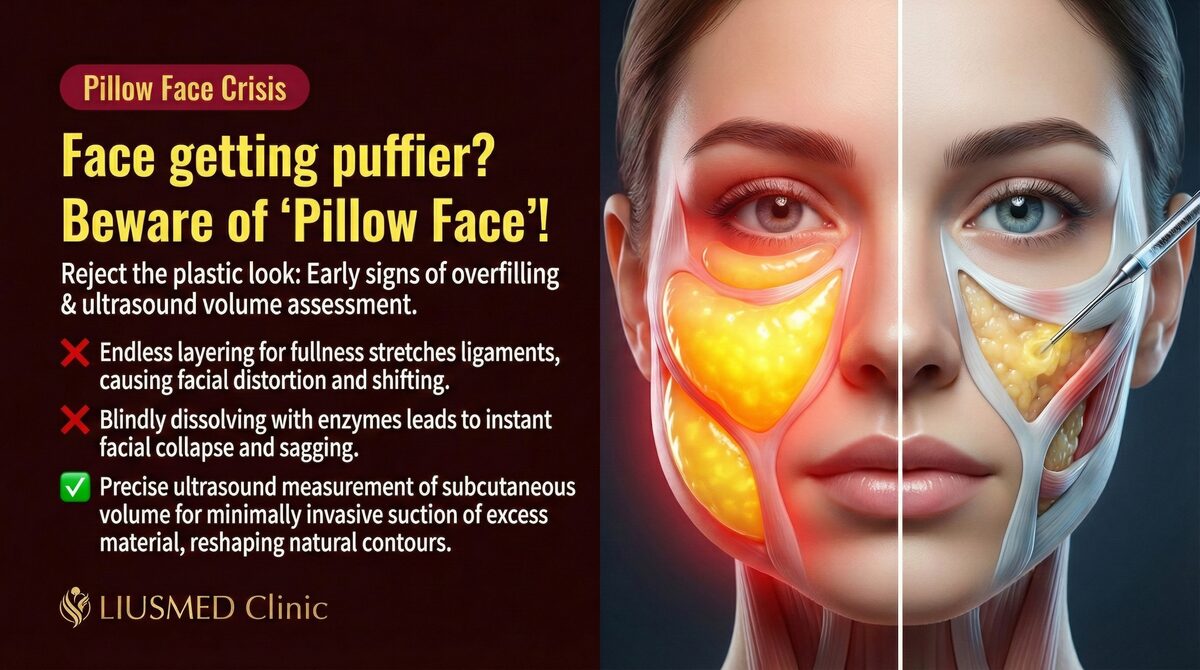
Key insight: The lump in Ellansé complications is fundamentally a fibrous capsule enclosing PCL microspheres — not a chemical property of PCL itself. Removal targets this capsule complex, not molecular dissolution of PCL. That is why "no enzyme" does not mean "no option."
2025 Exploration: Collagenase for Capsule Dissolution (Emerging and Contested)
In 2025, the Journal of Cosmetic Dermatology published a notable but still clinically contested approach: ultrasound-guided intralesional collagenase injection for Ellansé M nodules.
The proposed mechanism:
- Collagenase acts not on the PCL microspheres but attempts to break down the fibrous collagen capsule surrounding them
- Ultrasound precision aims to deliver collagenase into the capsule while limiting dispersion into surrounding tissue
- The original case series (only 3 patients) reported significant reduction of medium-sized Ellansé M nodules
This technique is not yet broadly accepted. A commentary published in the same journal raised explicit safety concerns about facial use — collagenase can cause vascular injury, severe bruising, and tissue necrosis, and the relevant collagenase product (approved only for cellulite) was withdrawn from the market in late 2022, making this an off-label application; the original authors replied defending the method using a recombinant collagenase and a larger case count.
Our position: Collagenase capsule dissolution is a direction worth watching, but the current evidence — a small case series and an expert debate — is not sufficient to recommend it as routine care. For mature, fibrotic nodules, this clinic continues to rely on the better-evidenced ultrasound-guided physical extraction.
Ultrasound-Guided Single-Pinhole Physical Extraction
For highly fibrotic nodules — particularly L- and E-type Ellansé, or cases where conservative management has failed — ultrasound-guided single-pinhole physical extraction is the most reliable approach:
- Ultrasound assessment: Map nodule position, depth, margins, and degree of fibrosis
- Local anaesthesia: Targeted anaesthetic to the treatment zone
- Single small entry point: One minimal incision under real-time ultrasound monitoring
- Precise extraction: Micro-cannula or needle aspiration of the fibrous capsule complex
- Immediate confirmation: Ultrasound confirms clearance of the target area
"See it to treat it safely" — ultrasound makes vessels, nerves, and capsule margins visible before the clinician acts, fundamentally changing the risk profile compared with blind manoeuvres.
How Ellansé Type Affects Difficulty
The molecular weight of PCL — and therefore the capsule maturity — differs by type:
← Swipe to see more →
| Type | Duration | PCL Mol. Weight | Capsule Thickness (clinical) | Conservative Response |
|---|---|---|---|---|
| S (1-year) | 12 months | Lowest | Thin, softens well | Relatively good |
| M (2-year) | 24 months | Moderate | Moderate | Collagenase investigational/contested (2025) |
| L (3-year) | 36 months | High | Thick, mature fibrosis | Limited; extraction indicated |
| E (4-year) | 48 months | Highest | Thickest | Limited; physical extraction often required |
For a detailed breakdown of how each Ellansé variant presents and is managed differently, see: Ellansé S, M, L — Complication Differences and Removal.
Ellansé Asymmetry After Injection: How Much Can Physical Revision Achieve?
What Causes Asymmetry
Asymmetry is the second most common reason patients seek revision after Ellansé, and its cause determines the correction strategy:
Technical causes:
- Unequal volumes between sides (a 0.1–0.3 ml difference can produce visible asymmetry)
- Depth inconsistency (a shallower side shows more immediate volumisation)
- Differential collagen induction: even with equal volumes, individual tissue variability causes unequal collagen production on each side
Material causes:
- During the CMC absorption phase (weeks 4–8), apparent volume changes unevenly, unmasking previously hidden differences
- Early PCL microsphere migration (facial expression or external pressure in the first month can shift product distribution)
Why "Add to the Deficient Side" Is Usually the Wrong First Move
With hyaluronic acid, adding to the thinner side is sometimes appropriate. With Ellansé, the logic is different:
Reasons to avoid immediate top-up of the deficient side:
- If the root cause is injection depth error, adding product to the same plane compounds the problem
- Collagen induction responses are unpredictable; an equal top-up may overshoot
- If early PCL migration caused the asymmetry, newly added product may migrate in the same way
The correct logic is subtraction first:
- Ultrasound bilateral comparison to confirm which side carries excess
- Precise extraction of the excess side
- 1–3 months of observation for tissue stabilisation
- Only then consider whether targeted augmentation of the deficient side is warranted
Key insight: The first question in asymmetry revision is never "which side needs more?" — it is "which side has too much, and where exactly is the excess?" Bilateral ultrasound delivers the objective answer that palpation cannot.
Expected Outcomes by Asymmetry Type
← Swipe to see more →
| Asymmetry pattern | Physical extraction prognosis |
|---|---|
| Focal excess on one side (clear nodule/cluster) | Excellent; visual symmetry often achievable |
| Diffuse excess (no discrete nodule, just more volume) | Good; targeted extraction reduces bulk |
| Differential collagen induction (no discrete excess) | Partial; extraction removes product but collagen production continues — monitor 3–6 months |
| Product migration cluster | Good once located under ultrasound |
Radiesse (CaHA) Calcification Nodules: What to Do
Clarifying "Calcification"
The phrase "Radiesse causes calcification" is widespread online, but it requires precision.
What CaHA actually is: Radiesse's active component is CaHA (calcium hydroxylapatite) microspheres — the same mineral found naturally in bone and teeth. When correctly injected, CaHA spheres are engulfed by macrophages and gradually metabolised, serving simultaneously as a collagen scaffold. This process is biodegradable and biocompatible by design.
What actually goes wrong:
- Superficial placement: CaHA spheres in the dermis, where macrophage clearance is insufficient, producing persistent palpable nodules
- Localised overinjection: High CaHA density in one area, exceeding clearance capacity, leading to a dense, well-defined aggregate
- Chronic granuloma: A minority of cases develop sustained foreign-body granulomatous reaction, producing a firm, warm, tender nodule
Why this matters for management: CaHA aggregates from injection errors are not pathological calcification in the clinical sense (not ectopic bone formation). Their management pathway — ultrasound-guided physical extraction — is correspondingly less radical than the term "calcification" might suggest.
Why Ultrasound Is Essential Before Any Intervention
CaHA microspheres are strongly hyperechoic on ultrasound — among the most sonographically visible of all injectable fillers. This property is an advantage for management:
- Precise nodule mapping: exact position, depth, and extent
- Inflammation assessment: a hypoechoic halo around the nodule suggests active inflammation, guiding whether to treat conservatively first
- Extraction monitoring: real-time confirmation of clearance
Plain radiograph (X-ray) can also demonstrate dense CaHA deposits, but provides no depth information and offers less diagnostic value than ultrasound for planning intervention.
Key insight: "It feels slightly firm" does not automatically require intervention. After ultrasound evaluation, many presentations turn out to be normal late-phase CaHA (no action needed), while some clinically inconspicuous cases reveal significant concentrated nodules requiring treatment. Imaging drives the decision, not palpation alone.
Does Sodium Thiosulfate Dissolve Radiesse?
A small number of off-label case reports describe sodium thiosulfate injection to chelate calcium from Radiesse deposits:
- Theoretically chelates calcium ions from CaHA
- No RCT evidence; only small case series
- Applicable only in a very narrow window: recently injected, uniformly distributed product before fibrous encapsulation has formed
- For mature, fibrotic CaHA nodules, the risk-benefit ratio is unfavourable
Ultrasound-guided physical extraction remains the best-evidenced approach for persistent Radiesse nodules. For a full breakdown of Radiesse calcium deposit management, see: Radiesse Complications and Calcium Deposits.
Radiesse Nodule Management: A Complete Pathway from Conservative to Surgical
Stage 1: Assessment and Classification (weeks 1–2)
Before any treatment, structured evaluation:
- Clinical history: injection date, anatomical zone, injector, symptom timeline
- Ultrasound evaluation: nodule location, fibrosis extent, rule out infection or vascular event
- Palpation correlated with imaging: consistency, mobility, fluctuance (suggesting fluid)
This stage determines which management branch to take; it cannot be skipped.
Stage 2: Conservative Management (the 3–6 month window)
Appropriate for:
- Nodules within 3 months of injection (fibrosis not yet fully mature)
- Ultrasound-confirmed ill-defined margins with inflammatory activity
- Mild symptoms (palpable only, no pain)
Conservative options:
← Swipe to see more →
| Approach | Regimen | Indicated when | Caveats |
|---|---|---|---|
| Warm compress + gentle massage | Daily | Early, mild | Clinician-supervised technique; wrong pressure worsens |
| Triamcinolone acetonide | Dilute 2.5–5 mg q4–6 weeks | Inflammatory, moderate firmness | Limit total injections; atrophy risk |
| Oral NSAIDs | Short course | Acute inflammation | Does not alter fibrosis |
| Sodium thiosulfate | Off-label | Very early, non-fibrotic only | Highly selective use |
Stage 3: Ultrasound-Guided Intervention
For conservative management failures at 3–6 months, or for clearly fibrotic nodules identified from the outset:
← Swipe to see more →
| Technique | Best indication | Expected outcome |
|---|---|---|
| Ultrasound-guided triamcinolone | Inflammatory type, moderate firmness | 50–80% reduction in most cases |
| Ultrasound-guided aspiration | Partially liquefied nodule on scan | Immediate volume reduction |
| Ultrasound-guided single-pinhole extraction | Dense fibrotic type, large nodule, repeated conservative failure | Most definitive clearance |
Stage 4: Open Surgical Excision (minority of cases)
Required only when:
- Nodule continues to enlarge beyond 6 months despite intervention
- Signs of chronic infection: erythema, warmth, pain persisting > 3 months; ultrasound shows fluid collection or air bubbles (biofilm signature)
- Nodule size is too large for complete clearance via single-pinhole access
Key insight: The majority of Radiesse nodules do not require open surgery. Ultrasound-guided minimally invasive removal bridges the gap between conservative management and open excision, covering most moderate-to-severe presentations.
Ellansé Complications: Staging, Timing, and Management Choices
Understanding the clinical stage of an Ellansé complication determines the correct management approach.
Acute Phase (0–4 weeks)
- Marked swelling, erythema, pain beyond normal post-procedure reactions
- Priorities: rule out vascular event (skin blanching, dusky discolouration, progressive necrosis → immediate emergency management) and infection (spreading erythema, fever)
- If confirmed inflammatory only: oral anti-inflammatories, cold compress; avoid heat and pressure
Early Fibrotic Phase (1–3 months)
- Palpable firmness with residual elasticity
- CMC largely absorbed; PCL microspheres in active fibrotic encapsulation
- Best conservative window: Triamcinolone and gentle massage are most effective here, before the capsule matures (intralesional collagenase has been explored at this stage but remains investigational and contested — see above)
- Start ultrasound evaluation to establish baseline
Chronic Fibrotic Phase (> 3 months)
- Well-defined, firm, low-elasticity nodule
- Ultrasound: hyperechoic nodule with thick fibrous wall
- Conservative efficacy limited; extraction indication becomes clear
- Delay compounds difficulty — capsule thickens with time
Delayed Immune Reaction (6 months to 2 years post-injection)
- Previously stable injection site develops new erythema and swelling
- Common triggers: viral illness (including COVID-19), vaccination, dental procedure
- Management: address immune trigger simultaneously; Triamcinolone ± antiviral/anti-inflammatory therapy
- Tends to respond better to Triamcinolone than chronic fibrotic nodules
Management Decision Summary
← Swipe to see more →
| Presentation | Stage | Key investigation | First-line approach |
|---|---|---|---|
| Swelling, erythema, pain | Acute | Rule out vascular event | Anti-inflammatory; urgent if vascular |
| Soft firmness, elastic feel | 1–3 months | Ultrasound | Triamcinolone + massage |
| Firm nodule, defined margins | > 3 months | Ultrasound | Guided extraction |
| Asymmetry | Any | Bilateral ultrasound | Subtraction from excess side |
| New flare (stable site) | > 6 months | Ultrasound + immune assessment | Treat trigger + Triamcinolone |
| Growing nodule + pain | Any | Urgent ultrasound | Rule out infection/biofilm; consider surgery |
For a more detailed examination of whether Ellansé can be removed and the biological basis, see: Can Ellansé Be Removed?
Ellansé vs Radiesse Revision: Side-by-Side Comparison
Both materials share the foundational principle: visualise before you intervene. The differences in material properties shape how each is managed.
← Swipe to see more →
| Dimension | Ellansé (PCL) | Radiesse (CaHA) |
|---|---|---|
| Dissolving enzyme | None (no PCL-specific enzyme) | None (thiosulfate off-label, limited) |
| Ultrasound visibility | Hyperechoic; locatable | Strongly hyperechoic; easiest filler to find |
| Main conservative option | Triamcinolone; collagenase (2025, investigational/contested) | Triamcinolone; warm compress |
| Fibrosis progression | Increases with type (S < M < L < E) | Depends on injection depth and volume |
| Extraction indication | L/E type; fibrotic; conservative failure | Dense fibrotic; 3–6 months conservative failure |
| Extraction technical notes | PCL spheres small; capsule must be captured | Strong US signal aids precision |
| Natural degradation | 1–4 years (type-dependent) | 12–24 months |
| Delayed immune reaction | Uncommon | Uncommon; COVID-era cases noted |
For the complete knowledge framework covering all collagen-stimulating fillers, see the pillar article: Collagen Stimulator Complications: Complete Overview.
FAQ
Will Ellansé lumps resolve on their own?
Early nodules within 1–3 months — soft, elastic, without defined margins — sometimes soften as CMC is absorbed, and gentle massage may help. But nodules that persist beyond 3 months with well-defined margins have a mature fibrous capsule; spontaneous resolution is unlikely. Waiting typically means a thicker capsule and harder management later. Ultrasound assessment followed by a planned approach is strongly preferable to indefinite observation.
Is Radiesse removal the same procedure as HA filler removal?
No. Hyaluronic acid has a dedicated enzyme (hyaluronidase) that chemically dissolves it — a drug injection. CaHA has no equivalent enzyme; physical extraction is the primary intervention. Both can be performed under ultrasound guidance, but the tools, technique, and tissue interactions differ. Interestingly, CaHA's strong hyperechoic signal makes Radiesse nodules among the easiest to locate precisely under ultrasound, which partially offsets the lack of chemical dissolution.
Will removing Ellansé cause sagging or hollowing?
Extraction is designed for precision — targeting the nodule complex while leaving surrounding tissue undisturbed. Ultrasound guidance keeps the procedure localised to the identified target. Post-procedure swelling is normal for several days. Actual tissue depression should not occur. In cases where the original injection created an overfilled appearance, extraction often restores a more natural contour. The real point is not merely "no sagging" but an even result — leaving no new dents or ridges. Removal done without enough control often turns a single lump into a whole uneven surface, which simply creates a different problem. The cross-disciplinary hand-feel Dr. Liu internalized over years of underarm rotary-blade surgery, liposuction and fat grafting, and the repair of failed liposuction is exactly what keeps the result even and precise — removing only what is needed, allowing for how expression moves the tissue. For the many patients who had material removed elsewhere yet were left uneven and came back for help, this is the difference that matters most.
When is open surgery necessary vs. minimally invasive extraction?
Open surgery is indicated for a minority of cases: large liquefied collections with chronic infection signs (> 6 months), confirmed biofilm with recurrent inflammation, or nodule volume that exceeds what single-pinhole access can clear. For the vast majority of moderate-to-severe nodules, ultrasound-guided single-pinhole physical extraction provides a definitive result without open incision.
Starting a Revision Assessment
For Ellansé or Radiesse complications — lumps, asymmetry, calcification, or recurring inflammation — revision planning always begins with ultrasound evaluation to characterise the nodule, rule out serious complications, and map a personalised treatment strategy.
No clinician can give you a meaningful answer before imaging. "See it to treat it safely" is not a slogan — it is the operating principle.
To schedule a revision consultation with Dr. Ta-Ju Liu: Book a Consultation
For an overview of the full filler revision service: Filler Revision Service
For the complete treatment methodology index: Treatment Overview
Editorial review: Dr. Ta-Ju Liu, Filler Revision Clinic