The Hidden Risk After Radiesse Injection: Why Does the Jaw Keep Getting Wider?
"My chin keeps getting wider, and I can feel hard lumps that weren't there before. My doctor says there's no enzyme to dissolve Radiesse — so what do I do?" At FILLER REVISION, Radiesse complications are among our most technically demanding cases because there is no pharmacological shortcut. Patients arrive after months or years of waiting for "natural absorption" that never happened, with jaw widening, palpable calcified nodules, and tissue fibrosis that steroids and massage cannot resolve. In our experience treating hundreds of CaHA (Calcium Hydroxyapatite) complications, ultrasound-guided minimally invasive extraction is the primary definitive solution when no dissolver exists.
Key Insight: At FILLER REVISION, we see this pattern regularly — while Radiesse is marketed as being absorbed within 12–18 months, clinical practice reveals that residual deposits and calcification are far from rare, particularly when large volumes are injected or placement depth is incorrect.
Radiesse Composition and Metabolism
Understanding CaHA Microspheres
The core component of Radiesse consists of CaHA microspheres approximately 25–45 micrometers in diameter. These particles share the same chemical composition as human bone and teeth minerals. Clinical safety studies of CaHA for soft tissue augmentation have documented its properties and complication profiles (Marmur et al., 2009). In theory, the body gradually breaks them down into calcium and phosphate ions for natural elimination.
← Swipe to see more →
| Component | Function | Expected Metabolism |
|---|---|---|
| CaHA microspheres | Structural support, collagen stimulation | 12–18 months (theoretical) |
| CMC (Carboxymethyl Cellulose) gel carrier | Immediate volume, microsphere distribution | 2–3 months |
| Neocollagen | Sustained volumizing effect | Persists but gradually diminishes |
Why Radiesse Cannot Be Dissolved (Brief)
Unlike hyaluronic acid fillers, Radiesse has no corresponding dissolving enzyme. Hyaluronidase is designed exclusively to cleave the molecular bonds of hyaluronic acid — it has zero efficacy against CaHA. See the dedicated section below for the full pharmacological explanation and how this compares to Ellansé and Sculptra.
Why Hyaluronidase Doesn't Work on Radiesse — And What Does
One of the most common — and costly — mistakes we see at FILLER REVISION is patients who were injected with hyaluronidase by a previous clinic in an attempt to dissolve a Radiesse lump. Months pass, the lump does not soften, the capsule continues to mature, and by the time the patient seeks a second opinion the fibrous tissue is denser and more difficult to extract than it would have been at month one.
The Pharmacological Mismatch
Radiesse is composed of two clinically distinct phases:
← Swipe to see more →
| Phase | Material | Behavior in Tissue |
|---|---|---|
| Microspheres (~30%) | Calcium hydroxylapatite (CaHA), 25–45 μm | Stimulate collagen, persist as palpable calcium particles for years |
| Gel carrier (~70%) | CMC (carboxymethylcellulose) gel | Resorbs over 2–3 months |
Hyaluronidase is a hyaluronic acid–specific hydrolase. It cleaves the β-1,4 and β-1,3 glycosidic bonds of HA chains. CaHA is a calcium phosphate mineral — chemically closer to bone than to a polysaccharide — and the CMC carrier is a cellulose derivative. Neither contains a hyaluronic acid backbone, so neither is a substrate for hyaluronidase. Injecting hyaluronidase into a Radiesse deposit is pharmacologically equivalent to injecting saline.
What Actually Remains in the Tissue
The CMC gel carrier resorbs within 2–3 months. What patients are feeling years later is not the original gel — it is the residual CaHA microspheres plus the fibrous capsule that has formed around them. Because the calcium particles cannot be enzymatically degraded and because the host capsule continues to thicken, the lump often feels harder, not softer, with time.
How This Compares to Other Biostimulators
← Swipe to see more →
| Filler | Particle | Carrier | Dissolving Option | Practical Removal |
|---|---|---|---|---|
| Radiesse | CaHA microspheres | CMC gel | None | Ultrasound-guided physical extraction |
| Ellansé | PCL (polycaprolactone) microspheres | CMC gel | None | Ultrasound-guided physical extraction |
| Sculptra | PLLA (poly-L-lactic acid) microparticles | No gel (reconstituted powder) | None | Physical extraction more diffuse; often steroid + extraction combined |
| HA fillers | Crosslinked hyaluronic acid | — | Hyaluronidase | Enzymatic dissolution |
The takeaway: all three biostimulators (Radiesse, Ellansé, Sculptra) share the same fundamental limitation — no enzyme exists to dissolve them. Sculptra's complication pattern tends to be more diffuse because there is no gel carrier and the PLLA powder disperses more widely; Radiesse and Ellansé both deposit as discrete microsphere clusters and are more amenable to localized extraction. For a deeper dive on Ellansé-specific patterns by formulation duration, see Ellansé S/M/L Complication Differences.
Why This Matters Clinically
When a general aesthetic clinic encounters a Radiesse lump and reflexively reaches for hyaluronidase:
- The patient pays for an injection with no pharmacological mechanism to help
- Several months are lost to "waiting to see if it works"
- During this delay the capsule continues to mature and the CaHA aggregates further
- By the time the patient arrives at a revision specialist, the extraction is technically harder
What Does Work
The practical options that actually address CaHA deposits are mechanical and supportive, not enzymatic:
- Ultrasound localization — CaHA appears as a distinctive hyperechoic (bright white) signal, making it among the easiest fillers to locate with imaging
- Micro-incision physical extraction — 1–2 mm pinhole entry, real-time ultrasound guidance, instrument fragmentation of the capsule and removal of the CaHA aggregate
- Intralesional steroid (selective use) — can soften an active inflammatory component, but has limited effect on already-calcified or fibrotic tissue, and carries skin atrophy risk with repeated dosing
- Time, with realistic expectations — waiting for "natural absorption" of CaHA is not a viable long-term strategy once a mature capsule has formed; the capsule itself persists even after microsphere degradation
Key Insight: If a clinician offers to "dissolve" your Radiesse with hyaluronidase, the conversation is over. Hyaluronidase has no mechanism of action on CaHA. The only definitive options for symptomatic Radiesse deposits are physical extraction or accepting the residual.
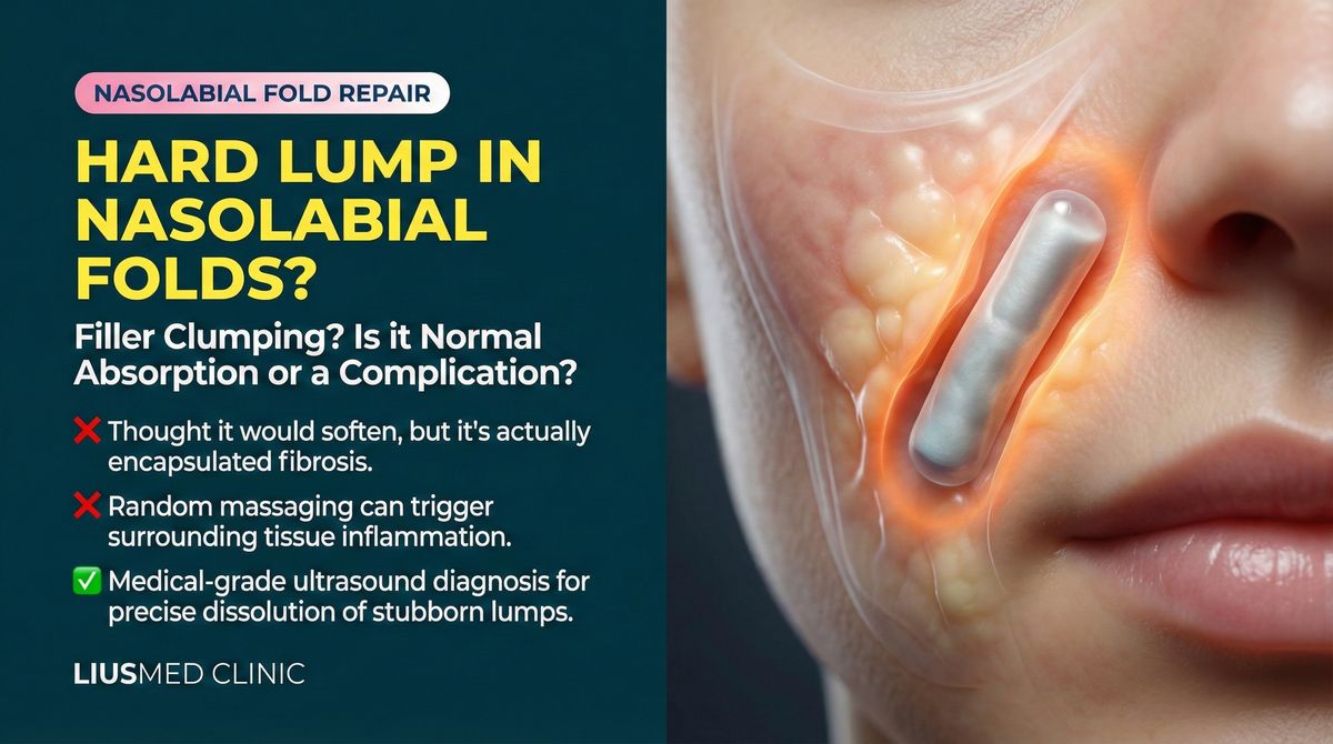
Common Radiesse Complications
Nodule and Lump Formation
CaHA microsphere aggregation within tissues can produce palpable hard lumps. Contributing factors include:
- Incorrect injection depth: Superficial placement allows microspheres to form visible clusters
- Excessive volume: Over-injection in a single site prevents even microsphere distribution
- Individual tissue response: Some patients mount a more aggressive fibroblastic reaction, accelerating capsule formation
- Poor local circulation: Slower microsphere metabolism leads to prolonged retention
Jaw Widening
The chin is one of the most common Radiesse injection sites, and jaw widening is a particularly distressing complication:
← Swipe to see more →
| Cause | Mechanism | Appearance |
|---|---|---|
| Filler spread | Microspheres disperse laterally along fascial planes | Chin appears squared from the side |
| Capsule formation | Fibrous tissue encases the entire deposit | Hard plate palpable on touch |
| Tissue fibrosis | Chronic foreign body reaction stimulates fibrosis | Tight, inelastic chin skin |
| Calcified aggregation | Microspheres cluster into larger calcified masses | Small stone-like feel on palpation |
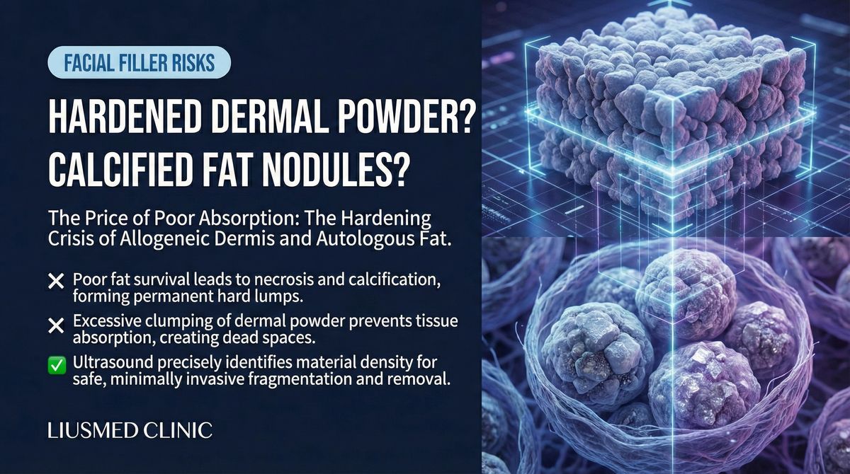
Tissue Fibrosis and Calcification
Long-term CaHA retention can trigger a cascade of pathological changes:
- Foreign body giant cell reaction: Immune cells attempt to engulf microspheres but cannot fully digest them
- Chronic inflammation: Persistent low-grade inflammation stimulates fibroblast activity
- Capsule formation: Fibrous tissue progressively encases the filler deposits
- Dystrophic calcification: Additional calcium deposits may form around retained microspheres
Limitations of Conventional Treatments
Massage and Observation
Many patients are told to "wait — Radiesse will absorb on its own." However, if palpable lumps or contour irregularities persist beyond 18–24 months post-injection, spontaneous absorption is unlikely. By this stage, the capsule has matured, and even if CaHA microspheres eventually degrade, the fibrous capsule will persist.
Steroid Injections
Intralesional steroid injections can temporarily manage inflammatory nodules, but carry significant risks:
← Swipe to see more →
| Potential Benefit | Potential Risk |
|---|---|
| Temporary anti-inflammatory effect | Skin atrophy and depression |
| Nodule softening | Hypopigmentation |
| Pain relief | Telangiectasia |
| — | Fat atrophy |
Steroids have limited effect on already-calcified or fibrotic tissue, and repeated injections accumulate tissue damage risk.
Surgical Excision
Traditional open surgical excision can remove Radiesse deposits, but performing open surgery on the face means larger incisions, greater scarring risk, and longer recovery. For patients who initially sought cosmetic enhancement, this is rarely the ideal option.
Ultrasound (Ultrasonography)-Guided Minimally Invasive Extraction: The "See Before You Treat" Strategy
Why Ultrasound Is Critical
Radiesse has a distinctive ultrasound signature — CaHA microspheres appear as hyperechoic (bright white) signals, making ultrasound the ideal tool for locating residual deposits.
← Swipe to see more →
| Ultrasound Capability | Clinical Value |
|---|---|
| Hyperechoic identification | CaHA clearly visible on ultrasound, precise localization |
| Depth and extent mapping | Quantifies residual filler distribution |
| Capsule assessment | Evaluates fibrous capsule thickness and extent |
| Vascular mapping | Avoids critical blood vessels |
| Real-time confirmation | Monitors extraction progress throughout the procedure |
The Extraction Procedure
Phase 1: Comprehensive Ultrasound Assessment
- Map all CaHA deposit locations, dimensions, and depths
- Evaluate capsule maturity and degree of fibrosis
- Plan optimal pinhole entry routes
Phase 2: Minimally Invasive Extraction
- Local anesthesia
- Pinhole incision (typically 1–2mm)
- Real-time ultrasound-guided approach to the target area
- Specialized instruments fragment the capsule and extract CaHA deposits
- Continuous ultrasound monitoring confirms extraction progress
Phase 3: Post-Procedure Follow-Up
- Light compression dressing for 24–48 hours
- One-week post-procedure assessment
- Follow-up ultrasound at 1 month and 3 months
Expected Outcomes
← Swipe to see more →
| Scenario | Expected Result |
|---|---|
| Single nodule | >90% removal in one session |
| Multiple nodules | May require staged treatment |
| Extensive fibrosis | Significant improvement; multiple sessions possible |
| Jaw widening | Contour gradually restores after removal |
| Injection over 3 years ago | Some CaHA may have degraded; capsule can still be extracted |
Key Insight: The goal of ultrasound-guided extraction is not to remove every microscopic CaHA particle, but to remove the principal deposits causing the clinical problem — restoring both appearance and texture to normal.
When No Dissolver Exists: The FILLER REVISION Approach
The fundamental challenge with Radiesse complications is irreversibility — unlike HA (Hyaluronic Acid) fillers, there is no enzyme, no medication, and no injection that can dissolve CaHA microspheres. Patients who reach FILLER REVISION have typically exhausted every non-surgical option: waiting for absorption that never comes, steroid injections that risk skin atrophy, and massage protocols that cannot break down calcified deposits. At FILLER REVISION, ultrasound-guided extraction addresses this directly. CaHA microspheres produce a distinctive hyperechoic signal on ultrasound, making them among the easiest fillers to locate with imaging. Our extraction protocol uses this visibility advantage — mapping every deposit, planning optimal pinhole entry routes, and extracting calcified material under real-time guidance while preserving surrounding tissue. For patients told their primary option is open surgical excision, this minimally invasive approach offers meaningful improvement without the scarring and prolonged recovery of traditional surgery.
Frequently Asked Questions
Will there be a depression after Radiesse removal?
Some degree of volume reduction is normal after extraction. Tissues remodel over 3–6 months. If volume restoration is desired, safe HA filler can be placed once healing is complete.
How long after injection can Radiesse be removed?
Extraction is technically possible at any time. Clinically, if significant residual symptoms persist beyond 18 months, active evaluation for extraction is recommended. Earlier treatment means thinner capsules and easier removal.
Will the extraction leave scars?
Pinhole incisions (1–2mm) heal with virtually invisible marks. Compared to traditional surgical excision, the cosmetic advantage of minimally invasive extraction is substantial.
Why does my Radiesse lump still get harder years later?
The CMC gel carrier in Radiesse resorbs within 2–3 months, but the CaHA microspheres themselves persist for years and cannot be enzymatically degraded. Around these residual particles, the host tissue forms a fibrous capsule as part of a chronic foreign body reaction. Over time the capsule continues to mature — collagen fibers cross-link, fibroblasts contract the surrounding tissue, and additional dystrophic calcium can deposit on the capsule wall. The clinical result is a lump that feels harder and more defined at year three than it did at year one, even though the original gel is long gone. This is why "wait for it to absorb" advice becomes less and less useful as time passes: the CaHA does not absorb on a timescale that matters, and the capsule actively becomes more difficult to break down. Ultrasound assessment can directly show capsule thickness and CaHA distribution, which is why we recommend imaging-based evaluation rather than another round of palpation alone.
Can Radiesse cause cosmetic issues even when it's "supposed to be absorbed"?
Yes — and this is one of the most common reasons patients arrive at FILLER REVISION. The marketing claim that Radiesse is "fully absorbed within 12–18 months" applies primarily to the CMC gel carrier and the initial volumizing effect. The CaHA microspheres themselves have a much longer residence time, often producing palpable particles, lateral spread along fascial planes, and contour irregularities long after the official "absorption window" has closed. Common late-onset issues include jaw widening from CaHA migration along the mandibular fascia, palpable calcified nodules in the chin and cheek, and tissue fibrosis around clustered microspheres. Because these problems develop slowly, patients often do not connect them to a Radiesse injection performed two or three years earlier. Ultrasound imaging makes the residual CaHA immediately visible as hyperechoic foci, which is usually the moment the timeline becomes clear.
Is Radiesse extraction safer than for HA filler?
Different, not strictly safer or riskier. With HA filler, hyaluronidase offers a non-surgical option for many uncomplicated cases — but once HA is encapsulated, the enzyme often cannot reach the gel and physical extraction is needed anyway. With Radiesse, there is no enzymatic shortcut at any stage; physical extraction is the practical definitive option from day one. The technical advantage with Radiesse is that CaHA produces a distinctive hyperechoic signal on ultrasound, making localization unusually clear — among biostimulators, Radiesse deposits are some of the easiest to image and target. The technical challenge is that mature capsules and aggregated calcium particles require careful instrument work to fragment without damaging surrounding tissue. In experienced hands using ultrasound guidance and 1–2 mm pinhole access, the procedure is well-tolerated; the safety profile depends primarily on operator experience and accurate imaging rather than on the filler material itself. A hybrid product like HArmonyCa (HA + CaHA) sits between the two: the HA component can be dissolved, but the CaHA component cannot, so it is only half-reversible — the residual CaHA still has to be physically extracted. See what to do about HArmonyCa nodules.
Do Not Let Residual Radiesse Control Your Appearance
If you've already tried treatment for Radiesse complications without success — or been told to simply wait for absorption that shows no sign of happening — FILLER REVISION specializes in exactly these cases. Our ultrasound-guided extraction is the definitive solution when no dissolver exists.
Further reading:
- Ellansé S/M/L Complication Differences
- Why Dissolving Enzymes Fail When Capsules Form
- Minimally Invasive Filler Lump Extraction Technique
- Lumps Appearing Years After Injection
About the Author
Dr. Ta-Ju Liu
- Current Position: Director, Liusmed Clinic
- Specialties: Minimally invasive surgery, filler complication repair, ultrasound-guided extraction
- Experience: 15+ years of clinical minimally invasive surgery; over 10,000 successful cases
- Philosophy: "Radiesse retention is more common than most realize. Patients should not have to choose between living with lumps and undergoing major surgery — ultrasound-guided minimally invasive extraction offers a third path."