Three months after a Sculptra treatment, a patient notices a firm nodule on the cheek. Six months later, after five rounds of steroid injections, the lump still hasn't resolved — and ultrasound reveals a dense fibrous capsule encasing the poly-L-lactic acid (PLLA) particles. This scenario is not rare: in a retrospective study of 130 patients, nodules occurred in 8.5% (Palm MD, Woodhall KE, Butterwick KJ, Goldman MP. Dermatol Surg. 2010;36(2):161-170. PMID: 20039924). Reported rates vary considerably between series, tracking the dilution volume and injection technique of the era (see "Why lumps form" below). One distinction matters first, though: a palpable nodule and a true foreign body granuloma are not the same thing. Genuine granuloma is far rarer — the literature puts it between 0.02% and 1% (Lemperle G, Gauthier-Hazan N, Wolters M, et al. Plast Reconstr Surg. 2009;123(6):1842-1863. PMID: 19483587) — and it is that subset that resists pharmacological treatment. Most nodules settle with conservative management; only a minority ever reach the point of needing removal.
This guide is for patients who have developed a hard lump, nodule, or granuloma after Sculptra, or who have been told their complication may be a granuloma. Understanding each step — from correctly identifying the nodule type, to knowing when steroid therapy is no longer the right tool — can prevent years of unsuccessful treatments.
For a broader overview of collagen-stimulator (PLLA / PCL / CaHA) complications, see the Collagen Stimulator Conditions Overview.
What to Do About a Sculptra Hard Lump: The First Assessment Steps
A "lump" and a "nodule" are the same finding named at two levels of precision. Lump is the word patients use for anything firm they can feel; nodule is the clinical term for a discrete, firm focus that persists on examination; granuloma is a tissue diagnosis, confirmed by histology or strongly suggested by the ultrasound findings described later in this guide. So wherever this guide says "nodule", it means the lump you can feel, and the table below sorts it by onset and behaviour.
A firm spot that can be felt but not seen after a Sculptra injection does not automatically indicate a problem. The biological mechanism of PLLA — sustained low-grade controlled inflammation that stimulates collagen — means some degree of early firmness is expected. The following table helps classify which category a lump falls into:
← Swipe to see more →
| Type | Onset | Characteristics | Recommended Action |
|---|---|---|---|
| Normal swelling | Days 0–7 | Diffuse swelling, no focal hard point, non-tender | Observe, gentle massage |
| Early small nodules | Weeks 1–4 | Palpable but invisible micro-particles, scattered | Active massage, observe for about 3 months |
| Delayed onset nodule (DON) | Week 4 to years later | Single or few firm, discrete nodules; may be visible | Specialist assessment required |
| True granuloma | Usually > 3 months | Hard, fixed, encapsulated, possibly tender | Active intervention needed |
| Infection / biofilm | Any time | Redness, warmth, swelling, purulence, significant tenderness | Seek immediate medical care |
More than half of small nodules that appear within the first three months resolve spontaneously with consistent massage. There is no need to panic. However, specialist assessment is appropriate if any of the following apply:
- The lump has not decreased in six or more months
- A visible surface elevation or asymmetry is present
- Tenderness is present and worsening over time
- Steroid injection provided temporary improvement followed by recurrence
Key point: "Palpable but invisible" does not equal urgent. The relevant question is whether the lump is shrinking, stable, or growing — and whether it is causing discomfort or visible change. Accurate classification prevents unnecessary early interventions and also prevents the opposite: waiting too long when true encapsulation has already begun.
Can Sculptra Nodules Be Removed? A Full Comparison of Non-Surgical and Surgical Options
The direct answer: Sculptra nodules can be treated, but the concept of "removal" is fundamentally different from that of hyaluronic acid (HA) fillers.
HA filler is a cohesive gel that can be dissolved by hyaluronidase (a dissolving enzyme) with high precision. Sculptra, by contrast, is a microparticle suspension — tens of thousands of PLLA particles dispersed throughout the tissue. There is no enzymatic agent that dissolves PLLA, and the dispersed nature of the particles makes "extraction of the whole mass" impossible in most cases. The four clinical options available are:
Option A: Intralesional Corticosteroid Injection (Triamcinolone Acetonide)
Mechanism: Direct injection of triamcinolone (Kenalog) into the nodule suppresses immune-mediated inflammation and reduces aberrant collagen deposition.
Appropriate when: The nodule is still in the inflammatory phase (tender, mildly red), has been present less than six months, and capsule formation is minimal.
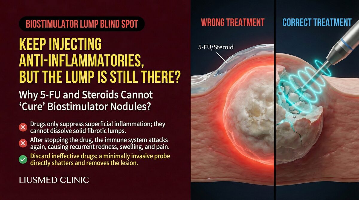
Limitations: Once significant fibrous encapsulation has occurred, the capsule acts as a barrier that prevents adequate steroid penetration to the PLLA particle core. Repeated injection attempts with no sustained improvement are a strong clinical signal that encapsulation is the underlying problem.
Option B: 5-Fluorouracil (5-FU) Combined with Corticosteroid
5-FU is an antineoplastic agent with anti-fibrotic properties in dermatological use, targeting the fibrous component of granulomas. In combination with steroids — which address the inflammatory component — the theoretical basis is sound, and published case series report positive outcomes for early-stage PLLA nodules.
The practical limitation is the same penetration barrier. For heavily encapsulated chronic PLLA granulomas, pharmacological agents continue to underperform regardless of the combination used.
For a detailed analysis of why 5-FU fails in encapsulated collagen-stimulator nodules, see: The Limitations of 5-FU for Collagen Stimulator Lumps
Option C: Ultrasound-Guided Physical Extraction
Under real-time high-frequency ultrasound guidance, a micro-cannula or aspiration instrument is advanced through a single 1–2 mm access point, directly targeting the encapsulated PLLA nodule. The capsule is mechanically disrupted and the loosened PLLA particles and fibrous tissue are aspirated.
Ultrasound-guided extraction rarely leaves a visible scar, because the only skin opening is a single 1–2 mm access point; what patients see at follow-up is usually a pinpoint mark. This is the main practical difference from open excision (Option D), where the incision has to be long enough to lift the granuloma out in one piece and therefore leaves a visible line. Dr. Ta-Ju Liu treats extraction quality as the second measure of a good result: the nodule is removed, and the aim is to leave the surface where it used to be even, without creating a new dip or step.
The core principle is "see it before you treat it" — ultrasound imaging allows the physician to identify nodule size, depth, and proximity to critical structures before any instrument enters the tissue, rather than working blindly.
Best suited for: Cases where steroids and/or 5-FU have been tried two or three times without clear improvement; clinical or ultrasound evidence of significant encapsulation; a single large, isolated, well-defined nodule; or patients seeking a more active approach.
Realistic expectations: In most cases, a significant reduction in nodule volume and resolution of visible surface irregularity can be achieved. Claims of "100% complete removal of all PLLA particles" are not clinically supportable — the microparticle nature of PLLA means total clearance confirmed on imaging has not been established in the literature. The goal is functional and aesthetic resolution, not a theoretically perfect scan.
Option D: Open Surgical Excision
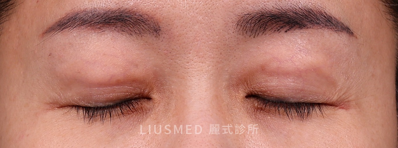
For anatomically localized confirmed granulomas — particularly in specialized areas such as the periorbital region — open excision remains a valid option. The primary tradeoff is the risk of visible scarring at the excision site. With advances in minimally invasive techniques, surgical excision is less commonly used as a first-line approach. At our clinic we do not perform surgical excision; granulomas that are extensive, widespread or deep are treated zone by zone over several sessions, still through pinholes of about 2 mm.
Key point: These options are not mutually exclusive. Early inflammatory nodules can reasonably begin with steroids. When encapsulation is established and drugs have repeatedly failed, continuing the same pharmacological approach produces diminishing returns, and minimally invasive removal is often the more reliable next step. Correct staging of the nodule — inflammatory versus fibrotic, encapsulated versus non-encapsulated — is the decision tree's starting point.
Sculptra Delayed Onset Nodules (DON): Why They Appear Months Later
"Why did my nodule only appear six months after the injection?" is a common clinical question. The answer lies in the distinct mechanism of delayed onset nodules (DON) compared with early technical nodules.
Early Nodule vs. DON: Two Different Problems
← Swipe to see more →
| Early Nodule (< 4 weeks) | Delayed Onset Nodule DON (> 4 weeks) | |
|---|---|---|
| Cause | Technical: uneven dilution, superficial injection, inadequate massage | Immunological: T-cell–mediated delayed-type hypersensitivity (Type IV) |
| Histopathology | Local PLLA particle aggregation | Foreign body granuloma — macrophages, epithelioid cells, multinucleated giant cells (MGC) surrounding PLLA particles |
| Texture | Small, scattered, may resolve spontaneously | Firm, fixed, discrete, single or few |
| Treatment response | Massage and observation usually effective | Variable pharmacological response; high failure rate after encapsulation |
The immunological mechanism of DON: PLLA particles degrade slowly over 2–5 years, continuously releasing antigenic signals during that period. In susceptible individuals, this sustained antigenic stimulus triggers a delayed-type hypersensitivity (DTH) response — immune cells cluster around PLLA particles and form granulomatous inflammation, inducing fibroblast proliferation and collagen deposition that progressively hardens into a fibrous capsule.
Reported triggers: Published case reports document a temporal association between systemic immune activation (viral illness, vaccination) and the onset of previously quiescent DON. This does not mean vaccination causes DON; rather, tissue containing dormant PLLA particles may develop focal immunological activity when the systemic immune system is broadly activated.
Why DON is particularly treatment-resistant: Once a fibrous capsule forms, it simultaneously blocks drug penetration to the PLLA core and shields the particles from phagocytic clearance — creating a pharmacological standstill where standard treatment agents cannot reach their target.
How Long Do Sculptra Nodules and Granulomas Last?
How long a Sculptra nodule lasts depends on which of the three categories it belongs to, and the ranges are very different. Early technical nodules that appear within the first three months are the short-lived group: more than half resolve with consistent massage, usually within the massage and re-assessment window shown in the table above. Delayed onset nodules and true granulomas follow a much longer course. Lemperle and colleagues (2009, PMID 19483587) describe foreign body granulomas after injectable fillers as appearing several months to years after injection and, without treatment, growing to the size of beans, remaining virtually unchanged for some years, and then resolving spontaneously. Put in patient terms, an untreated PLLA granuloma is measured in years, and its eventual resolution follows the slow degradation of the particles themselves (roughly 2–5 years, as described above).
Treatment does not reliably shorten that course once a capsule has formed. In the Brazilian series of 55 collagen-biostimulator complications reported by Ianhez and colleagues (2024, PMID 38693639), nodules were the most common presentation (89.1%) and most appeared late (60.0%, beyond one month); despite saline, hyaluronidase, diluted corticosteroids and energy-based devices, only five cases showed complete resolution. That is also the pattern Dr. Ta-Ju Liu sees in the Sculptra patients who reach Filler Revision: the great majority have already had steroids, 5-FU, or both, often over months to years, and the nodule is still palpable. A mature capsule does not dissolve on its own; the lump may soften slightly over years but usually remains palpable until it is physically removed. The question to settle is which category you are in and whether a capsule has formed, and that is what the ultrasound assessment in the next section answers.
PLLA Granuloma Risk Factors: Not Random, and Traceable
With thousands of successful Sculptra treatments worldwide, why do granulomas occur in some patients and not others? Identifiable risk factors exist on both the technical and patient-specific side.
If you have not been injected yet, the four technical factors below double as a pre-treatment checklist. Ask your injector which plane the product will be placed in, how many millilitres each vial is diluted in, what massage schedule you are expected to follow, and how much volume is planned per session. Checking all four does not remove the risk entirely, but each one is a variable the injector controls, and most of the technical risk described in this guide comes from these four.
Injection Technique Factors
1. Superficial injection depth PLLA should be deposited in the subcutaneous fat or supraperiosteal plane. Intradermal or superficial subdermal injection places PLLA particles in a microenvironment with high immune cell density and poor vascular supply, significantly increasing granuloma incidence.
2. Insufficient dilution Current guidelines recommend diluting one vial of Sculptra in ≥ 7–9 mL of sterile water. Earlier protocols using only 3–5 mL produced higher particle concentrations in tissue. Many legacy cases of PLLA granuloma — including cases presenting years after injection — trace back to the older dilution regimen.
3. Inadequate post-injection massage The "5-5-5 massage rule" (5 minutes, 5 times daily, 5 days post-injection) is designed to distribute PLLA particles evenly and prevent focal aggregation. Insufficient massage creates locally concentrated particle clusters that increase granuloma trigger probability.
4. Excessive injection volume per session Particularly in the deep zygomatic region, high single-session volumes increase regional granuloma risk regardless of other technical factors.
Patient-Specific Factors
- Autoimmune predisposition: Patients with a history of Sjögren's syndrome, systemic lupus erythematosus, or rheumatoid arthritis have a higher baseline risk of PLLA-induced granuloma and should receive detailed pre-procedure counseling.
- Pre-existing biofilm: Areas with a history of prior filler injections may harbor subclinical biofilm. PLLA injection into these areas can precipitate infection-triggered granuloma — a distinct subtype requiring antibiotic biofilm treatment before the nodule can be adequately reduced.
- Thin skin and minimal subcutaneous fat: Periorbital and neck regions have less fat buffer between skin and any injected PLLA, placing particles closer to the dermis where immune activity is higher.
The sites most often reported with Sculptra nodules are the hands and the cheeks. In the 130-patient series by Palm and colleagues (2010, PMID 20039924), the treatment areas with the highest nodule incidence were the hands (12.5%) and the cheeks (7.2%), and almost all nodules were palpable rather than visible. The cheeks matter because they usually receive the largest volumes; the hands, the periorbital area and the neck matter because there is little fat between the skin and the deposited PLLA. Both patterns come back to the same two variables discussed above, volume per site and how close the particles sit to the dermis.
Key point: PLLA granuloma formation is rarely fully random. In most cases, one or more identifiable risk factors can be traced retrospectively. This is an argument for thorough pre-procedure assessment — immune history, prior filler history, tissue thickness — rather than treating Sculptra as a universally "low-risk" collagen stimulator.
For further reading on 5-FU treatment limitations in collagen-stimulator granuloma: Collagen Stimulator Lumps and 5-FU Treatment Failure
After Steroid and 5-FU Failure: Advanced Revision Strategies
For context on why repeated steroids fail to resolve encapsulated PLLA nodules, see: Why Sculptra Lumps Don't Respond to Repeated Steroid Injections
The most common clinical path for unresolved Sculptra nodules is: initial steroid injection → partial temporary improvement → recurrence → second steroid injection → diminishing response. If this cycle has repeated two or three times without clear improvement, continuing the same approach is unlikely to produce a different outcome. The strategic reframe at this point is: assess the nodule's actual anatomical and biological state, then choose a tool that matches it.
Step 1: Ultrasound Assessment of Encapsulation
High-frequency ultrasound (15–22 MHz) is the key evaluation tool before any further intervention. An encapsulated PLLA granuloma presents with:
- A hyperechoic, densely structured capsule border
- Internal echogenic foci representing PLLA particle aggregates
- Increased perinodular blood flow on Doppler, indicating active inflammation
Clinical interpretation: When capsule thickness exceeds approximately 1 mm and nodule diameter exceeds 8 mm, drug penetration through the capsule is substantially reduced. At this stage, physical disruption of the capsule structure typically yields more reliable results than additional injection attempts.
Step 2: Ultrasound-Guided Mechanical Capsule Disruption and Aspiration
Under real-time ultrasound guidance:
- Fine-needle entry into the capsule, with a small volume of saline or steroid solution to soften the capsule interior
- Micro-cannula insertion through a single 1–2 mm access point to mechanically disrupt the capsule structure
- Negative-pressure aspiration of fragmented PLLA particles and fibrous tissue
- Real-time ultrasound verification of residual volume
In most cases, this achieves substantial reduction of residual nodule volume and resolution of visible surface irregularity. As noted earlier, confirmed total clearance on imaging is not a realistic endpoint given the microparticle nature of PLLA — the clinical goal is relief of symptoms and aesthetic improvement.
Step 3: Post-Procedure Management and Follow-Up
- Week 1: Antibiotic coverage if any signs of infection or biofilm are present
- Months 3–6: Ultrasound follow-up to assess residual PLLA signal and local inflammatory activity
- If symptomatic residual nodule persists: evaluate need for second-session intervention
Emerging Options (Evidence Still Accumulating)
JAK inhibitors: There is accumulating clinical evidence that JAK inhibitors (targeting the JAK-STAT immune signaling pathway) can provide partial improvement in injection-related granulomas unresponsive to conventional treatment. Standardized protocols are still being established; Dr. Liu will discuss this option based on each patient's individual case and current literature.
Frequently Asked Questions
My Sculptra has gone wrong and I have lumps — what should I do first?
Classify the lump before treating it. Diffuse swelling in the first week and scattered micro-nodules at 1–4 weeks usually need nothing more than observation and massage. A single firm nodule appearing four weeks or later is a delayed onset nodule (DON); one that is hard, fixed and encapsulated beyond three months is handled as a true granuloma. Redness, warmth, swelling, purulence and marked tenderness point to infection or biofilm — seek medical care immediately. The tool that settles this is high-frequency ultrasound, not another blind injection. If steroids have already failed twice, that is the point to reassess the strategy rather than repeat it.
Will Sculptra lumps go away on their own?
More than half of the small nodules that appear within the first three months resolve with consistent massage, so early firmness is not a reason to panic. But a lump is unlikely to resolve by waiting if it has not shrunk after six months or more, if there is visible elevation or asymmetry, if tenderness is worsening over time, or if a steroid injection shrank it only for it to come back. Those patterns usually mean a fibrous capsule has formed — and the longer encapsulation continues, the harder later intervention becomes.
How long do Sculptra nodules and granulomas last?
Early technical nodules usually settle within weeks to a few months of consistent massage; true granulomas can persist for years. Lemperle and colleagues (2009, PMID 19483587) describe foreign body granulomas after fillers as remaining virtually unchanged for some years before resolving spontaneously, in step with the slow degradation of the PLLA particles. Once a fibrous capsule has formed, medication rarely shortens that course: in the 55-case series by Ianhez and colleagues (2024, PMID 38693639), only five cases resolved completely despite several treatment types. An encapsulated nodule that is still there at six months is unlikely to disappear by waiting, which is why ultrasound staging comes before any further injection.
Steroid injections aren't clearing my Sculptra nodules. What now?
Shrink-then-rebound is the classic encapsulation pattern: the steroid calms the inflammation around the nodule but never penetrates the capsule to reach the PLLA core, so the nodule returns once inflammation restarts. On ultrasound, when capsule thickness exceeds roughly 1 mm and nodule diameter exceeds 8 mm, drug penetration is substantially reduced and further blind injections have little left to offer. The reasonable next step is ultrasound-guided mechanical capsule disruption with minimally invasive removal: entry through a single 1–2 mm access point, disruption of the capsule, then aspiration of the fragmented PLLA particles and fibrous tissue. Because PLLA is a microparticle suspension, the goal is relief of symptoms and of visible surface irregularity — not a claim of clearing every particle.
Will treating the granuloma reverse Sculptra's collagen-stimulating effect?
Treating a granuloma does not undo Sculptra's cosmetic benefit. A granuloma represents pathological over-fibrosis, not the desired controlled collagen induction — removing it addresses the problem tissue, not the treatment result. In cases where granuloma involves the majority of the treated region, the overall aesthetic assessment should be discussed individually with the physician.
Can I ask to have all the Sculptra removed?
Because PLLA is a microparticle suspension rather than a cohesive mass, there is no "total extraction" equivalent to removing an HA filler. Symptomatic nodules and granulomas can be substantially cleared. Asymptomatic PLLA particles distributed in soft tissue do not require — and realistically cannot be — completely removed. The assessment focus should be on whether a specific symptom or visible concern is causing distress, not on achieving zero residual PLLA.
The lump got smaller after steroid injection but came back. What does that mean?
This is the classic encapsulation pattern: steroids reduce perilesional inflammation and the nodule appears to shrink transiently, but the drug has not penetrated the capsule to address the PLLA core. When inflammation restarts, the nodule rebounds. The cycle of partial shrinkage and recurrence is a clinical indicator that the current treatment modality is insufficient and escalation should be considered.
Does every delayed onset nodule need treatment?
Not necessarily. If a nodule can be felt but not seen, causes no discomfort, and shows no progression, observation may be appropriate — some DON cases resolve as PLLA gradually degrades over several years. Active intervention is indicated when there is visible surface elevation, progressive enlargement, significant tenderness, or failure to respond to pharmacological treatment.
If your Sculptra nodule has been present for more than six months and has not responded to two or three rounds of steroid treatment, waiting further is rarely in your interest — the longer encapsulation persists, the more challenging any subsequent intervention becomes.
Dr. Ta-Ju Liu uses ultrasound-guided minimally invasive techniques and has managed multiple cases of treatment-resistant PLLA granuloma with significant encapsulation. Visit the Filler Revision Clinic service page to understand the full assessment and treatment process, then schedule a consultation to discuss your individual case.
Dr. Ta-Ju Liu | Filler Revision Clinic — fillerrevision.com