"I felt a few hard lumps after AestheFill, my doctor said to wait and inject one more time, but I've injected three or four times and they're still there." "Should I keep injecting, or just have them dealt with?" — In clinic, nodules from AestheFill (poly-D,L-lactic acid, known in some markets as the "elf needle") are something we take over often, and the place patients get stuck is rarely "can this nodule be treated." It's "which step am I supposed to be on right now."
Treating AestheFill nodules is actually a ladder. From the most conservative option, massage, all the way down to minimally invasive removal, every rung exists for a reason — and every rung has a ceiling. The trouble is that many people circle around the "drug injection" rung or two for half a year without moving down, while the tissue keeps getting irritated. This article opens up the whole ladder so you know what each rung is doing, and when it's time to move down.
Key insight: The first mistake in managing AestheFill nodules is usually not "choosing the wrong method" — it's "staying on one rung too long." When a method has been tried two or three times and the lump hasn't shrunk, that's usually your body telling you it's time to move to the next rung.
Why AestheFill nodules need a "ladder" approach
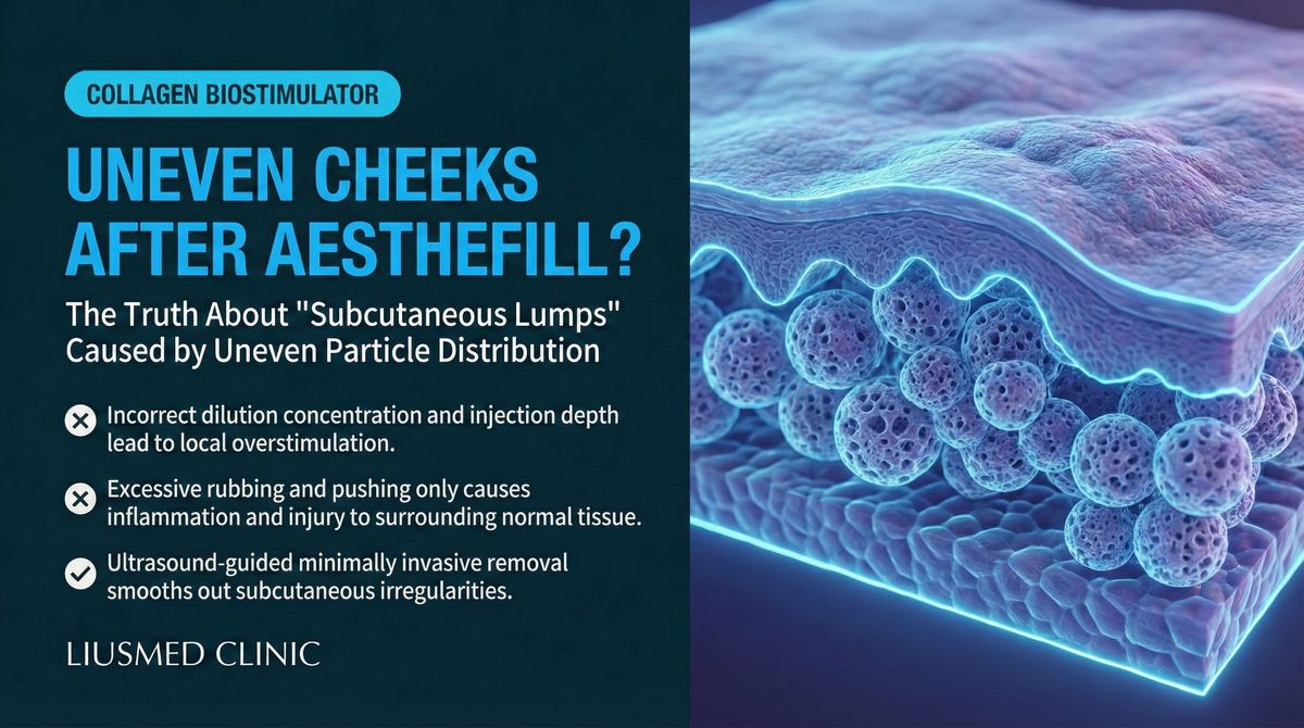
AestheFill's main ingredient is PDLLA (poly-D,L-lactic acid) microspheres, which work by stimulating your own collagen to create a filling effect — it's a type of collagen stimulator. It's a cousin of Sculptra's PLLA (poly-L-lactic acid), but PDLLA particles are more uniform and the texture is softer. Nodules tend to form when the injection is too concentrated, the layers are uneven, or an individual's collagen response is unusually strong.
But before doing anything, one thing matters more than choosing a method — first figure out which kind of nodule this is. A 2025 guideline in the Journal of Cosmetic Dermatology, compiled by two authors (PMC11956123), puts exactly this at the front: first rule out the things that look like nodules (skin calcification, epidermoid cysts, even a few skin tumors), then use ultrasound to see clearly.
By time of onset, nodules roughly fall into three categories:
← Swipe to see more →
| Time of onset | Likely nature | How the approach differs |
|---|---|---|
| Days to weeks after injection | Leans infectious | Treat the infection first (antibiotics, drainage if needed), don't rush to massage |
| 3 months to years | Delayed hypersensitivity | May need anti-inflammatory treatment, not simple physical release |
| 6 to 24 months | Granulomatous nodule | Often needs an anti-inflammatory strategy such as corticosteroids |
Classification directly decides which rung you should start on. If an inflammatory nodule is massaged or subcised first, it can actually get worse; a simple non-inflammatory aggregate is the one suited to the physical-release-to-removal line.
Key insight: Even when they all feel like "a hard lump," the cause underneath might be material aggregation, fibrous encapsulation, or an immune reaction — and the logic for treating each is completely different. That's why we insist on "see it before you treat it": ultrasound first is the starting point of the whole ladder, not one option among many.
For what each filler material looks like on ultrasound and how to tell them apart, see our piece on the ultrasound filler material comparison.
Rung 1: Massage and observation
For an early, non-inflammatory small nodule, massage is a reasonable first step. The 2025 guideline above also lists "vigorous massage" as a non-invasive initial approach. The idea is to use external force to help disperse the locally aggregated microspheres.
Its role: lowest threshold, lowest risk. Its ceiling is just as clear — once a mature fibrous capsule has formed around the nodule, external force struggles to penetrate, and massage just makes you feel reassured while the lump itself barely moves.
When to leave this rung: a few weeks of massage plus observation with no change, or a nodule that instead grows harder or larger — that's when you should stop relying on kneading.
Rung 2: Saline lavage
The next rung down is local lavage with normal saline. The concept is to use a relatively gentle method to add local hydration, dilute, and try to loosen the aggregated material — gentler than injecting an irritating drug straight away.
Its role is the in-between option of "doing a bit more than massage, but not yet injecting a drug," suited to the stage where massage hasn't worked but you're not yet at the point of needing a drug. Its ceiling is the same: for an old nodule already wrapped in dense fibrosis, lavage alone can only do so much.
Rung 3: Subcision plus drug injection
Further down is subcision (releasing the fibrous adhesion under the skin with a needle) combined with drug injection. The drugs often mentioned include 5-FU (5-fluorouracil, an antimetabolite that suppresses fibroblasts), corticosteroids (intralesional injection to suppress inflammation), and some experimental options.
Two things have to be said honestly about this rung:
First, the literature itself says it's of limited effect. That 2025 PDLLA guideline, in describing subcision with drug injection, used very blunt wording: this approach "is not particularly effective and can lead to additional setbacks." That's not our claim — it's what the authors who compiled the guideline wrote based on their clinical experience. It echoes what we see in clinic over and over: for nodules that are already mature and fibrotically stable, repeated injections are often a lot of effort for little reward. For why 5-FU stalls, see collagen stimulator nodules: what to do after 5-FU fails.
Second, regarding collagenase (an enzyme that breaks down collagen), we do not offer this here. In 2025 there was indeed a tiny experimental study (PMC12038313, just 3 patients, 10 nodules) that tried using collagenase to dissolve Ellansé (PCL) nodules, with apparent reduction and no adverse events observed — but this is off-label, experimental use, the sample is far too small, and recurrence and long-term safety remain undefined; the authors themselves called for larger studies. It cannot be treated as proof that "AestheFill is solved," and it does not replace physical removal. For the details and limits of that study, see can collagenase dissolve Ellansé nodules?
Key insight: The biggest trap on the drug-injection rung is the "maybe one more shot will fix it" mindset. When a professional guideline plainly says the approach is of limited effect, and you've injected several times and are still in the same place, the signal is fairly clear: the problem may not be the number of injections, but that this nodule is no longer suited to being solved by a needle.
Rung 4: Radiofrequency / ultrasound energy
Further down the ladder, some try energy devices such as radiofrequency (RF) or ultrasound-type energy. The idea is to use heat to soften the PDLLA material and speed up its breakdown.
This direction is worth knowing about, but the evidence has to be stated clearly: what's published is only a single case report (PMC11743236, one 42-year-old woman with bilateral tear-trough PDLLA nodules), in which monopolar RF over two sessions was described by the authors as resolving the nodules within 24 hours, with no recurrence at the time. This is the lowest tier of medical evidence, and the authors themselves note it was the first attempt to use energy devices for these nodules and that more research is needed to establish protocols and safety.
In other words: RF for PDLLA nodules is an interesting, possibly promising direction — but it is nowhere near a validated standard approach. Treating it as "worth a cautious try in the right case, but not something to pin hopes on" is closer to reality.
Rung 5: When the ladder reaches the top — traditional surgical excision vs. ultrasound-guided minimally invasive removal
When the earlier rungs have all been tried and the nodule stubbornly remains, the ladder reaches its final rung: taking the material out.
Tellingly, that same 2025 guideline ends its final rung with exactly this — "in cases where the nodule persists, surgical excision may be required." In other words, even the literature cataloguing chemical and drug methods concedes that stubborn nodules ultimately need physical removal. The only difference is — how you take it out.
← Swipe to see more →
| Traditional surgical excision | Ultrasound-guided minimally invasive removal (our final solution) | |
|---|---|---|
| Access | Larger incision | 1–2mm pinhole |
| Can you see it | Mostly by experience and touch | Real-time ultrasound guidance, removing as you watch |
| Surrounding tissue | Larger area removed alongside | Precise separation along the nodule's border |
| Scarring risk | Higher | Pinhole heals, less visible |
| Goal of removal | Take the nodule out | Not just out, but cleanly and evenly |
This is also where we differ most from most approaches. For us, the deciding factor in removing an AestheFill nodule is never "whether it can be removed" — it's taking it out cleanly, taking it out evenly, precisely removing the aggregated material without leaving new irregularity on the face. Ultrasound here plays the role of "eyes": confirming the nodule's location, depth, and relationship to nearby vessels and nerves before the needle goes in, then removing as you watch in real time, getting the aggregated material out as cleanly as possible while preserving what should be preserved.
For the overall philosophy of this physical-removal route, and the causes of AestheFill lumps and the ultrasound removal detail, see repairing AestheFill lumps and uneven PDLLA complications, and you're welcome to learn about our filler repair service.
Which rung should I start on?
Not everyone has to start at rung 1 and climb all the way down. In practice, where you should enter depends on the nature of the nodule, how long it's been there, and what's been done before. The starting point for that judgment is always ultrasound.
What ultrasound can tell us, in terms a patient can follow: is this material aggregation, or fibrous encapsulation? Is it in a shallow or deep layer? Is there an important vessel beside it to avoid? Those answers decide whether you're suited to trying conservatively downward or to considering removal directly. In other words, the ladder isn't there to make you suffer through every rung — it's there to help you and your doctor judge "where you are now, and where the next step goes."
For telling nodules apart and when to seek care, see is my lump filler, granuloma, or scar?
When should I stop injecting and consider removal?
This is the question most often asked in clinic, and the one that most deserves a real answer. The following situations usually mean the payoff from continuing down the injection path is already low:
- The same method (massage, drug injection) has been tried more than three times with no clear improvement
- The skin has started to thin or atrophy after corticosteroids, but the nodule is still there
- The nodule is already over six months old and the fibrosis has stabilised
- The nodule is in a shallow layer, clearly affecting facial contour, and you no longer want to wait for it to degrade on its own
One thing to be clear about: minimally invasive does not mean painless. Removal is done under gentle pain-relief anaesthesia, with the doctor talking to you in real time and able to pause and adjust at any moment, keeping discomfort as low as possible — but it is still a procedure, not a no-sensation experience. We would rather tell you this honestly than trade your peace of mind for a promise of "feeling nothing."
AestheFill nodules don't disappear by waiting, and circling around the first few rungs only keeps the tissue irritated. If you've already tried massage and injected several times without improvement, you're welcome to come in with your history and imaging — we'll use ultrasound to see clearly first, then decide together which rung you should stay on, or whether to move toward removal. Book a consultation →
About the author
Dr. Ta-Ju Liu
- Current position: Director, Filler Revision Clinic
- Expertise: Extreme minimally invasive surgery, filler complication repair, ultrasound-guided extraction
- Experience: Over 15 years of clinical minimally invasive surgery, with more than 10,000 minimally invasive cases
- Philosophy: "In treating AestheFill nodules, what matters most isn't rushing to act — it's seeing clearly first, then deciding which rung of the ladder to move to. When injections keep failing, ultrasound-guided minimally invasive removal offers certainty — and the value of that certainty lies in taking the material out cleanly and evenly."
Frequently Asked Questions
Do AestheFill nodules have to be removed? Can I just wait for them to go away?
Not necessarily. An early, non-inflammatory small nodule can indeed start with massage and observation, and PDLLA material itself gradually degrades over time. The catch is the cost of "waiting": if the nodule is already mature and has formed a fibrous capsule, waiting could mean several years with your appearance affected throughout. Whether and when to remove should be decided with your doctor based on ultrasound assessment and your own tolerance for time — not a blanket "just wait" or "operate immediately."
I've already injected 5-FU and corticosteroids several times and the lump is still there — should I switch methods?
Quite possibly. A 2025 guideline on PDLLA nodules plainly states that subcision with drug injection "is not particularly effective and can lead to additional setbacks." When the same drug method has been tried more than three times with no clear improvement, or corticosteroids have been injected until the skin starts to atrophy while the nodule remains, it usually means the problem isn't the number of injections — it's that this nodule is no longer suited to being solved by injection, and it's worth an ultrasound assessment and considering the removal rung.
I've heard collagenase can dissolve AestheFill nodules — do you do that?
We do not offer collagenase injection here. At present, using collagenase to dissolve these collagen-stimulator nodules rests only on a tiny experimental study (3 patients), it is off-label use, and recurrence and long-term safety remain undefined — the study authors themselves called for larger research. It cannot be treated as proof that "AestheFill nodules now have a drug that solves them," and it does not replace physical removal. Our approach is ultrasound-guided minimally invasive removal, which offers certainty.
Can radiofrequency or ultrasound energy really clear AestheFill nodules?
The evidence is still very limited. What's published is only a single case report — one patient's bilateral tear-trough nodules resolving after monopolar RF. This is the lowest tier of medical evidence, and even the study authors say it was a first attempt that needs more research to establish protocols and safety. So RF / ultrasound energy can be seen as a "cautious try in the right case" in-between option, but it shouldn't be treated as a validated standard approach — nor should it become a reason to keep putting off treatment.
How does ultrasound-guided minimally invasive removal differ from traditional surgical excision?
The biggest difference is "being able to see" and "taking it out cleanly and evenly." Traditional surgical excision mostly relies on experience and touch, with a larger incision and a larger area removed alongside, so the scarring risk is higher. Ultrasound-guided minimally invasive removal goes through a 1–2mm pinhole, removing as you watch under real-time ultrasound and separating precisely along the nodule's border. For us, the point isn't just taking the material out — it's taking it out cleanly and evenly, without leaving new irregularity on the face. That's the real deciding factor in treating AestheFill nodules.