Your finger slides across your cheek while you wash your face, and it stops. There is something there now. Hard. It wasn't there before.
Over the next few days you will probably do the same thing on repeat: look in the mirror, press it, compare it with the other side, search "hard lump after filler." The answers you find will contradict each other. Some say it will go away on its own, some say dissolve it immediately, some say surgery. That is exactly how the anxiety builds.
Let me say the most important thing first. When you feel a lump, the right move is not to treat it — it is to first figure out what it is. Residual filler, a foreign-body granuloma, and scar tissue can feel very alike, but their causes differ, their course differs, and the right method for each is completely different. Treating a granuloma as if it were ordinary filler — rubbing it, topping it up with another injection — is often where a small problem turns into a big one.
This article cannot give you a definitive diagnosis through a screen; the final answer for any lump needs imaging. But it can do two things. First, it can use a few clues you can observe right now to help you sense which category you lean toward. Second, it can tell you which situations mean "stop waiting, see a specialist."
Start With These Three Self-Check Questions
Before you decide on a next step, observe the lump itself, calmly. The three questions below need no equipment. You can do them right now.
Question 1: Is it painful, red, or warm?
Press gently with the pad of your finger and pay attention.
- Not painful, not red, normal skin temperature: this leans "non-inflammatory." It is more likely residual filler, an encapsulation (a fibrous capsule the body wraps around foreign material), or a settled scar.
- Tender when pressed, flushed at the surface, warmer than the surrounding skin: this leans "inflammatory." This type deserves more caution — it may be an inflammatory nodule, an infection, or an immune reaction in progress.
Inflammatory does not automatically mean severe, but it does mean your body is reacting to this thing right now, which leaves less room to simply watch and wait than the non-inflammatory type does.
Question 2: Does it move, or is it stuck in place?
Place your finger on the lump and push gently in different directions.
- Moves, with a fairly clear border: more like an independent ball of filler or a capsule.
- Stuck deep and won't move, blurred border, blending into the surrounding tissue: be more careful. A lump with an unclear border is more likely to involve a tissue reaction, fibrosis, or a granuloma, and these carry a high risk if you handle them yourself.
Question 3: When did it appear?
This question is often overlooked, but it carries a lot of information. Think back: did this lump appear within a few days of the injection, or did it surface months later?
- Within days of injection, right where the needle went in: more likely related to how that session's filler was distributed or spread unevenly.
- A long time later, even more than half a year afterward, appearing suddenly: in medicine this is called a "delayed-onset nodule," and its meaning is completely different from an early lump. The next section is devoted to it.
Key point: Tenderness, mobility, and timing — no single clue settles it, but together they let you roughly tell which side of "inflammatory vs non-inflammatory" you fall on, and that is the dividing line that decides whether you can keep observing or how quickly you should be seen.
Inflammatory vs Non-Inflammatory: The Two Forks in the Road
Put the answers to those three questions together, and most lumps fall onto one of two paths.
← Swipe to see more →
| Clue | Leans non-inflammatory | Leans inflammatory |
|---|---|---|
| Tenderness | None or mild | Clearly tender |
| Color and warmth | Normal | Flushed, warm |
| Time of appearance | Early after injection | Any time, including delayed |
| Texture and border | Clearer, movable | Blurred, possibly tethered |
| Common correlates | Residual filler, encapsulation, settled scar | Inflammatory nodule, infection, foreign-body granuloma |
To be clear, this table shows a tendency, not a diagnosis. There are plenty of gray zones in practice — a long-stable piece of residual filler can suddenly flare after a cold or a dental procedure, crossing from non-inflammatory to inflammatory. So its purpose is to help you judge how urgent things are right now, not to slap on a final label.
If your lump sits clearly on the inflammatory side — especially if it is getting more painful, more red, or spreading — treat that as a signal to be seen rather than to keep waiting.
Delayed-Onset Nodules: Why Does It Surface Half a Year Later?
This is what confuses many people the most: everything was fine after the injection, so why does a lump appear out of nowhere so much later?
These "delayed-onset nodules" actually have a basis in the literature. A 2024 retrospective study in Frontiers in Microbiology analyzed 61 patients with delayed or late filler complications and found that nodules appeared on average about 16.2 months after injection, with a range stretching from 3 months all the way to 6 years. The study also observed that the trigger for these delayed reactions is often not the injection itself but something that happens afterward — an acute infection, another illness, a shift in the menstrual cycle, even mental stress can be the spark.
The researchers pointed to a "biofilm" (a thin bacterial film clinging to the filler surface) as one key risk factor. It sits there quietly until some change in the body wakes it up, at which point it can drive late swelling and nodules. This also explains a common frustration: oral medication relieves it at the time, but symptoms return once you stop — because the root has not been removed, only suppressed.
It is worth noting that this is a single-center, retrospective study with a limited sample, so it cannot be used to draw conclusions about your individual case. But it at least gives a reasonable explanation for "why so late," and it flags one thing: a delayed-onset nodule is usually not something that rubbing and waiting will resolve, because there may be an active reaction behind it that needs handling. For a fuller picture of how biofilm causes recurrent swelling, see our article on biofilm and recurrent filler swelling.
Filler, Granuloma, Scar: How They Fundamentally Differ
With the self-check done, the next step is understanding what these three things you are trying to tell apart actually are. They can all feel like "a hard one," but they are very different at the core.
- Residual filler: the material you were injected with is still there, or the body has wrapped it in a layer of fibrous capsule (encapsulation). It is not necessarily inflamed; the problem is that its position, shape, or texture bothers you, or it blocks a follow-up you want to do.
- Foreign-body granuloma: a chronic immune reaction to a "foreign body." The body treats the filler as an invader and surrounds it layer by layer with immune cells, forming a hard nodule. Its core is the immune reaction, not the material itself. To understand how this immune mechanism switches on, read further in how granulomas form.
- Scar tissue: the fibrotic mark left after tissue repair. It can come from the needle entry, a past procedure, or scarring after repeated inflammation. It is usually no longer "active," but its texture is harder than normal tissue.
The tricky part with all three is this: they need different — sometimes opposite — directions of treatment. Residual filler may be suited to located, physical extraction; the immune reaction of a granuloma has to be understood and controlled first; a stable scar usually does not need, and is not suited to, being approached with "remove the filler" logic. Acting before you even know which one it is makes it easy to go in the wrong direction.
Key point: "They all feel like a hard lump" does not mean "they are all handled the same way." Filler, granuloma, and scar are three fundamentally different things, and telling them apart is the first step of correct treatment, not a step you can skip.
Why Rubbing It or Topping It Up Yourself Often Makes It Worse
When people are anxious, they badly want to "do something." But for a lump, the two most common self-rescue moves often backfire.
Rubbing hard. Many assume a lump can be massaged away. But if it is a granuloma or an inflammatory nodule, mechanical irritation can actually make the inflammation more obvious; if it is filler, rough rubbing can push it into a layer or position it should not be in. We discussed this myth in full in can massage break up a filler lump?.
Another round of dissolver, or just topping it up to cover it. This is another common mistake. Dissolver (which should only be used after a doctor's in-person assessment) works on hyaluronic acid (HA), but if your lump is not HA at all — if it is a collagen stimulator, a permanent filler, or already a granuloma or scar — dissolver is not only ineffective, it can delay the treatment that is actually needed. As for "adding another injection to cover the unevenness," that is layering one more thing on top of a problem you haven't figured out, making it harder to sort out later. We cover the cumulative cost of repeated blind dissolving in the cumulative damage of repeated dissolving.
Put simply: before you know what the lump is, any "active treatment" is a gamble, and the cost of gambling wrong is usually paid by your face.
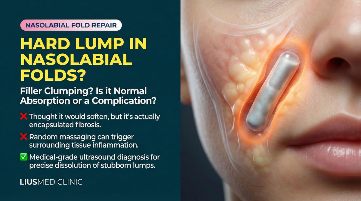
How Ultrasound Settles It, and When to See a Specialist
Every self-check above can only help you triage; none of it can give you the final answer. What settles it is imaging.
There is a core idea in filler revision: you can only treat safely what you can see. High-frequency ultrasound shows what is under the skin in real time and distinguishes its position, layer, and nature. The literature supports this too. A 2018 study in Skin Research and Technology (11 subjects) described the ultrasound features of different lesions: foreign-body granulomas tended to be oval, with blurred, irregular borders and small high-echo spots visible inside, while plain filler deposits were mostly anechoic with sharp, regular borders. This is a small-sample study and cannot guarantee any single case, but it makes one practical point: residual filler and granuloma leave traceable differences on imaging — differences your fingers cannot feel and only the eye can see on a screen.
In other words, what ultrasound answers are the very questions you care about most: is this filler? If so, which layer, does it move, has it been wrapped in a capsule? If it is not filler, does it look more like an inflamed granuloma or an already-stable scar? Only once it is seen clearly can you talk about "what to do next." For how far ultrasound can clarify things when the material is uncertain, see identifying an unknown filler.
As for when to stop watching and go straight to a specialist, here are some clear signals:
- The lump is getting more painful, more red, or spreading.
- A delayed-onset nodule appears — a lump that surfaces long after injection.
- Medication helps while you take it, but symptoms return the moment you stop.
- You aren't sure what material was injected in the first place, or more than one was used.
- The lump has a blurred border and won't move, or it affects your expression or appearance and keeps bothering you.
If any one of these fits, observing on your own is no longer advisable. Seeing it clearly sooner usually leaves you with more options, and with less drastic ones.
A Final Word
Feeling a lump is frightening — that is an entirely normal reaction. But what I want you to take away is a calmer order of operations: observe first, distinguish first, figure out what it is first, and only then talk about how to treat it — rather than the reverse, rushing to treat something you do not yet recognize.
Dr. Ta-Ju Liu has focused for many years on filler complication repair and has seen, in clinic, many cases pushed further out of reach by "just rub it" and "just add another syringe." If a lump is troubling you and you can't tell whether it is filler, granuloma, or scar, rather than searching yourself into a deeper panic, let imaging answer it. You are welcome to describe your situation through an online consultation, or to learn how our filler revision sees clearly under ultrasound guidance before deciding the next step. You can also find a closer match to your situation in our conditions overview.
This article is health education and cannot replace an in-person medical diagnosis. The literature cited is for research reference; individual samples are limited and do not represent every case. The final assessment and management of any lump still requires a case-by-case judgment after an in-person consultation and imaging.