A lot of patients sit down and, before asking about treatment, let out a sigh: "I can feel it, but my doctor told me there's nothing there."
They're not here to argue. Most of them feel that lump on the side of the nose every time they wash their face, press it and it even moves, go back to where they were injected to ask, and get brushed off with "there's nothing, you're overthinking it." After a while, some start to wonder if they really are being neurotic.
Honestly, I've seen this too many times. And almost every time I start with the same sentence: if you can feel it, you're usually not overthinking it. No one has shown it to you yet.
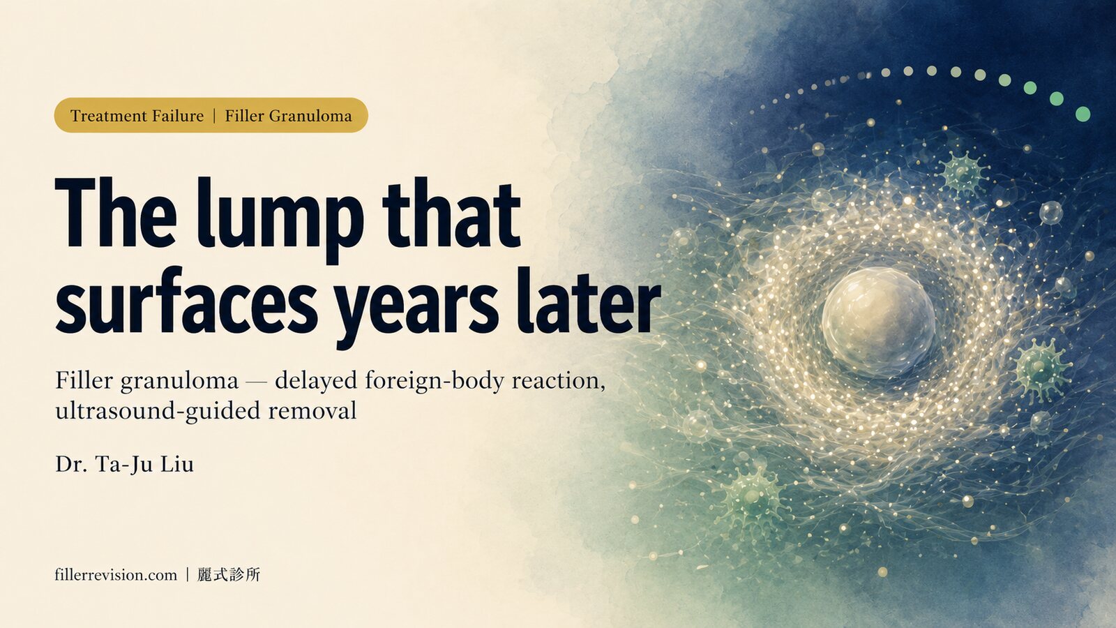
Why "I can feel it" gets answered with "there's nothing there"
Let's be clear about one thing. When the doctor who injected you says "nothing's there," most of the time he isn't lying to you. He just had two tools in hand at that moment: his eyes and his fingers.
The naked eye reads the surface. Once the swelling settles and the nose looks acceptable from outside, you genuinely can't see anything. Fingers can feel a lump, but they can't tell which skin layer it sits in, or whether it's retained filler, fibrotic scar, or normal cartilage. The nose magnifies that gap. The subcutaneous space is small and the structures are stacked tightly, so a modest piece of retained material hidden inside is hard to call by touch alone.
So "I feel it but I'm told it's nothing" is almost never a dispute about whether something is there. It's a question of whether the right tool was used to look.
Key insight: Palpation and the naked eye have a ceiling. To answer objectively whether there's residue in your nose, you don't need two opinions arguing. You need to image it.
What high-frequency ultrasound actually sees
The thing that can actually settle this is high-frequency ultrasound. It isn't the kind of scan used to look at your abdomen at a health check; the probe runs at a much higher frequency, built to look at the few millimetres of superficial skin structure, with enough resolution to tell filler, vessels and tissue layers apart.
It tells me three things touch can't.
First, which layer the residue is in and how big it is. On ultrasound, retained filler often appears as a hypoechoic lesion (dark on the scan), with measurable borders, depth and size. A very typical picture is a hypoechoic patch on one side of the nasal bone with the other side clean. Compare left to right, and the lump you've been feeling is finally seen.
Second, roughly what material it is. Different fillers have different echo signatures. Hyaluronic acid (HA) is usually a fairly uniform hypoechoic mass; biostimulators behave differently. Polycaprolactone (PCL, the main ingredient in Ellansé), for example, has been described in the literature as showing hyperechoic (bright on the scan) spots with comet-tail artifacts. The echo pattern, together with your history, helps estimate which class you were injected with, and that directly affects whether it can be dissolved and how it should be handled.
Third, its relationship to the vessels. The nose carries a relatively high risk of vascular complications, so whether the residue hugs a branch of the dorsal nasal artery is something to see clearly before doing anything.
This isn't theory. A 2024 study in J Cosmet Dermatol by Bravo and colleagues showed that under real-time high-frequency ultrasound guidance, nasal filler position and vessel course can be seen clearly, lowering vascular risk. A 2022 case report is even more direct: a patient with delayed-onset midface nodules was only identified as having deep PCL deposits thanks to 20 MHz ultrasound. The report says plainly that without imaging, she might have been put through repeated, ineffective superficial injections for nothing.
Palpation and the naked eye vs high-frequency ultrasound
← Swipe to see more →
| What you want to know | Palpation + naked eye | High-frequency ultrasound |
|---|---|---|
| Is anything actually there | Can feel it, but can't be sure | Objectively shows a lesion or not |
| Which layer, how deep | Can't tell | Measures layer and depth |
| Roughly what material | A guess | Estimated from echo pattern |
| Relationship to nasal vessels | Invisible | Mapped before removal |
| Whether it's safe to remove | No certainty | Decided after seeing it |
If you want a fuller picture of how ultrasound tells fillers apart, see how ultrasound identifies filler.
You have to see it to treat it safely
I say one thing to patients a lot: you have to see it to treat it safely. That's not a slogan. In the nose it's hard reality.
The vessels in the nose are fine and densely packed, so blind aspiration or blind scraping carries particular risk here. Operating without imaging is like working next to a vessel with your eyes closed. Seeing the residue clearly, which layer, how big, against which vessel, is itself the precondition for safety, not an optional extra. On the risks of extracting without ultrasound, I've written a piece specifically on the danger of blind extraction.
Key insight: "I feel it but I'm told it's nothing" usually means no imaging was done, not that there's truly nothing. Seeing the residue clearly on ultrasound first is where everything starts, and the floor for safety.
From "confirmed it's there" to "removed cleanly"
Once ultrasound confirms residue, then we talk about treatment.
If it's early, small-volume HA that hasn't encapsulated yet, dissolving with hyaluronidase (the enzyme that breaks down HA, used only after an in-person physician assessment) can be considered. But if the HA has already encapsulated (the body has wrapped the material in a fibrous capsule), or it's a biostimulator with no dissolving agent at all, repeated dissolving usually does nothing, and physical removal is the more direct route. Which materials can be dissolved and which can only be removed is laid out in the nose filler decision matrix.
Our ultrasound-guided removal follows the same principle: first map the residue's position, depth and its relationship to branches of the nasal artery on high-frequency ultrasound, then remove the material under image guidance through a very small entry point. The whole procedure uses gentle pain-controlled local anaesthesia, so the doctor and patient can talk in real time and pause to adjust at any point.
To be honest about it: removal does not come with a "guaranteed 100% clean" claim. Biostimulators, CaHA (calcium hydroxylapatite, the main ingredient in Radiesse) and permanent materials can adhere to tissue over the years, so the complete-clearance rate varies with material, time and degree of adhesion, clinically often around 80–90%. What we aim for is to greatly reduce the residue while keeping the nose contour even, without leaving new bumps.
A real example
I had a patient whose lump on the side of her nose had been with her for a full fifteen years. Over those years she asked several doctors and got wildly different answers, one of which was that line: "there's nothing here."
One sweep of ultrasound, and the hypoechoic patch on the right side of her nasal bone was perfectly clear, in exactly the spot she'd been feeling for fifteen years. What finally let her breathe out wasn't what anyone had said back then. It was that she could finally see, with her own eyes, that it really was still there. The full story is in this case.
If you want the complete picture of how retained nose filler is repaired, start with the retained nose filler and lumps overview.
FAQ
Q: I can feel it, but my doctor says there's nothing. Who do I believe? A: You don't have to guess. Palpation and the naked eye have limits; a single high-frequency ultrasound can objectively show whether there's residue in your nose and which layer it's in. Rather than trading opinions, see it clearly.
Q: Can high-frequency ultrasound tell exactly what I was injected with? A: It can estimate, not declare with certainty. Different materials have different echo signatures; HA, biostimulators and permanent fillers each look different on imaging, and combined with your history, that estimates the likely class, which then guides whether to try dissolving or remove directly.
Q: The residue has been there for over a decade. Can it still be dealt with? A: Yes. Residue doesn't disappear on its own just because time has passed; as long as ultrasound can locate it, removal can be assessed. The length of time isn't the deciding factor. Precise localisation and clean, even removal are.
Q: Can removal guarantee it's completely cleared? A: We don't use "guaranteed 100%." Biostimulators and permanent materials can adhere to tissue over time, so the complete-clearance rate varies by individual situation, clinically often around 80–90%. The goal is to greatly reduce residue and leave the nose contour natural and even.
See it clearly first, then decide
A lump you can feel on your nose, dismissed as "nothing," and what you actually want is simple: an objective answer. That answer isn't on anyone's lips. It's in the image.
See it clearly first: which layer, how big, against which vessel, then talk about whether and how to treat. If you're stuck at this step, you're welcome to use an online personalised assessment or book a consultation, where Dr. Ta-Ju Liu can help confirm what's actually in your nose with high-frequency ultrasound.
References
- Bravo BSF, de Melo Carvalho R, Elias MC, et al. Nasal filling guided by high frequency ultrasound: Reducing risks. J Cosmet Dermatol. 2024. (Real-time high-frequency ultrasound locates nasal filler and vessels to reduce risk.)
- Shekarriz P, Shojaee P. Ultrasound-assisted management of filler-related complications: Report of a successful treatment of delayed-onset nodules related to polycaprolactone-based filler. Clin Case Rep. 2022. (20 MHz ultrasound identifies the echo signature of deep PCL deposits; without imaging, repeated ineffective superficial injection is likely.)
Editorial Review: This article is educational information, not individual medical advice. Whether retained nose filler is present and whether to remove it must be decided case by case after an in-person physician assessment and ultrasound evaluation. Actual treatment and outcomes vary by individual.