"I Just Wanted It Removed—How Did Things Get Worse?"
"The lump is gone, but now I have a dent and my lip is numb." At FILLER REVISION, we see patients with this exact story every week — people who underwent blind aspiration or curettage at another clinic and ended up with damage worse than the original lump. Over 40% of our revision cases involve repairing harm caused by unsighted extraction attempts.
The problem was not a lack of skill. The problem was that the practitioner could not see. Without ultrasound or other imaging guidance, the practitioner inserts a needle, cannula, or curette into the tissue, relying on tactile feedback alone. On the face — densely populated with nerves, vessels, and delicate structures — this blind manipulation can produce consequences far more severe than the original complication.
What Are Blind Aspiration and Blind Curettage?
Blind Aspiration
Inserting a needle or cannula into tissue without imaging guidance, attempting to withdraw filler material through suction. The practitioner relies entirely on palpation and tactile feedback to locate the filler.
Blind Curettage
Inserting a curette or specialized instrument into tissue without imaging guidance, attempting to scrape out filler material and surrounding capsule tissue. This also relies entirely on touch and clinical judgment.
← Swipe to see more →
| Technique | Guidance Method | Precision | Risk Level |
|---|---|---|---|
| Blind aspiration | None—tactile only | Low | High |
| Blind curettage | None—experience only | Low | Very high |
| Ultrasound (Ultrasonography)-guided aspiration | Real-time imaging | High | Low |
| Ultrasound-guided micro-extraction | Real-time imaging + precision instruments | Very high | Lowest |
Five Critical Risks of Blind Aspiration and Curettage
Risk 1: Unable to Confirm Material Type
Without ultrasound, the practitioner cannot confirm the filler material before the procedure. Different materials have different textures, tissue adhesion patterns, and optimal removal strategies. Silicone extraction is entirely different from HA (Hyaluronic Acid) extraction, and blind manipulation cannot distinguish between them.
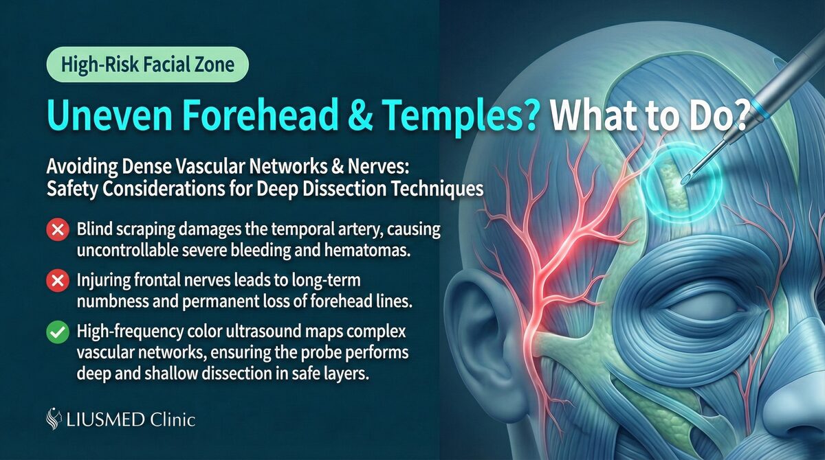
Risk 2: Nerve Damage
The face has an extremely dense distribution of motor and sensory nerves. Blind needle or curette movements can damage these nerves, causing:
- Localized numbness or sensory disturbance
- Facial muscle weakness or asymmetry
- Mouth drooping
- Permanent nerve injury
Risk 3: Vascular Injury
The facial vascular system is complex with numerous anatomical variations. Blind manipulation can puncture critical vessels, leading to:
- Significant bleeding
- Deep hematoma
- Skin necrosis (if supply arteries are compromised)
- Vision impairment (in extreme cases)
Risk 4: Excessive Removal of Normal Tissue
One of the greatest problems with working blind is the inability to distinguish the boundary between filler and normal tissue. During blind curettage, the practitioner may inadvertently scrape away normal fat and connective tissue, causing:
- Severe tissue depression
- Irreversible volume loss
- Irregular surface contours
Risk 5: Incomplete Removal and Recurrence
Paradoxically, blind procedures can also result in insufficient removal. Because the full extent of the filler cannot be visualized, the operator may miss residual material, leading to persistent lumps that require further treatment.
Key Insight: At FILLER REVISION, we see this fundamental contradiction play out in the patients who come to us after blind procedures: to avoid damage, the practitioner operates conservatively — but conservative operation leaves material behind. To achieve thorough removal, more aggressive technique is needed — but aggressive technique without visual guidance damages normal structures. Without imaging guidance, this contradiction cannot be resolved.
Common Sequelae We See After Blind Curettage
We regularly see patients in our clinic who seek help after undergoing blind curettage elsewhere. Common sequelae include:
- Irregular surface contours: Caused by uneven removal of normal tissue
- Skin tethering to deep tissue: Scar tissue pulls the skin down to deeper layers
- Residual lumps on the opposite side: Blind curettage only addressed what could be felt
- Post-operative infection: Open wounds increase infection risk
- Persistent numbness: Sensory nerves inadvertently damaged
Key Insight: The damage from blind curettage is sometimes harder to repair than the original problem. A filler lump can potentially be removed through precise minimally invasive surgery, but the tissue defects and scar adhesions caused by blind curettage multiply the complexity and difficulty of repair by several fold.
The FILLER REVISION Approach: Repairing What Blind Procedures Left Behind
Many patients arrive at FILLER REVISION not with their original filler problem, but with the aftermath of a blind extraction attempt — irregular contours, tissue adhesions, residual lumps, and nerve damage. Our revision protocol begins with comprehensive ultrasound mapping to assess what remains: residual filler, scar tissue, areas of over-removal, and the condition of surrounding structures. This detailed imaging allows us to plan a precise repair strategy — removing any remaining material while preserving healthy tissue. Because we can see every tissue layer in real time, we avoid the exact trap that caused the initial damage: guessing where the filler ends and normal tissue begins. For patients who have already suffered one blind procedure, this image-guided approach is not optional — it is essential.
Ultrasound Guidance: The Fundamental Shift from Blind to Seeing
High-resolution ultrasound has completely transformed how filler complications are managed. It provides more than just "better positioning"—it represents an entirely different treatment philosophy: do not begin any intervention until you can clearly see all the structures involved.
What Can Ultrasound Visualize?
- Precise filler location and depth
- Material characteristics (different materials show different echogenic signatures)
- Degree and thickness of encapsulation
- Surrounding vascular and nerve pathways
- Real-time position of surgical instruments
Ultrasound-Guided Minimally Invasive Extraction Process
- Pre-procedure scanning: Creates a complete "map" marking filler locations and structures to avoid
- Real-time guidance: Continuous ultrasound monitoring throughout the procedure confirms instrument positioning
- Precise extraction: Under visual confirmation, only filler material and necessary capsule tissue are removed
- Intraoperative verification: Real-time confirmation of clearance completeness ensures nothing is missed and nothing is over-removed
- Post-procedure follow-up: Ultrasound monitoring of recovery
For more detailed information on the minimally invasive extraction technique, see: Filler Lump Extraction Technique Explained.
Why Traditional Debridement Is Unacceptable on the Face
In certain medical contexts, debridement is necessary and effective—for example, removing necrotic tissue from deep wounds. But applying the same blind debridement logic to facial filler complications is an inappropriate transplantation of general surgical concepts into a field that demands fine-detail precision.
The face is unique because:
- Every millimeter of tissue can affect appearance
- Symmetry requirements are extremely strict
- Nerve and vessel density is far higher than elsewhere on the body
- Patient sensitivity to outcomes is extremely high
In this environment, "roughly knowing where the material is" is not sufficient. You must "precisely know where the material is, and what surrounds it."
How to Choose the Right Treatment Approach
If you are facing filler complications that require intervention, consider the following when choosing a treatment approach:
- Does the practitioner use ultrasound for pre-procedure assessment?
- Is real-time imaging guidance used during the procedure?
- Can the practitioner identify the filler material type and exact location before treatment?
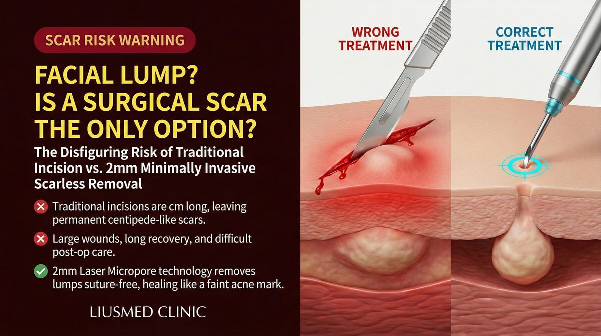
- Is the treatment performed through a tiny pinhole or does it require a surgical incision?
We recommend starting with a comprehensive ultrasound evaluation to clearly understand your situation before making treatment decisions. Schedule a consultation and let us use a visible approach to find the safest solution for you.
Conclusion
If you have already undergone a blind aspiration or curettage that left you with worse contours, numbness, or residual lumps, FILLER REVISION specializes in repairing exactly these cases. Our ultrasound-guided approach ensures that every step is performed under visual confirmation — so your revision does not repeat the mistakes of the first attempt. Your face deserves to be seen before it is treated.
Frequently Asked Questions
I had filler removed at another clinic and now I have a dent and numbness. Is this common, and can it be repaired?
This is a story we see regularly. Over 40% of our revision cases involve repairing harm caused by blind aspiration or curettage done without ultrasound guidance. FILLER REVISION specializes in exactly these cases — we begin with comprehensive ultrasound mapping of residual filler, scar tissue, and areas of over-removal, then plan a precise repair that removes remaining material while preserving healthy tissue.
What exactly are blind aspiration and blind curettage, and why are they risky on the face?
Blind aspiration inserts a needle or cannula to withdraw filler by suction, while blind curettage inserts a curette to scrape out filler and capsule tissue — both without any imaging, relying entirely on touch. On the face, which is densely packed with nerves, vessels, and delicate structures, this blind manipulation can produce consequences far more severe than the original complication.
Why does removing more filler blindly risk leaving a dent, while removing less leaves lumps behind?
Working blind creates an unresolvable dilemma. To avoid damage the practitioner operates conservatively — but conservative operation leaves material behind, causing residual lumps. To remove thoroughly, more aggressive technique is needed — but without visual guidance that aggressive technique scrapes away normal fat and connective tissue, creating dents and irreversible volume loss. Without imaging guidance, this contradiction cannot be resolved.
Is damage from a blind procedure harder to fix than my original filler problem?
Often, yes. A filler lump can potentially be removed through precise minimally invasive surgery, but the tissue defects and scar adhesions caused by blind curettage multiply the complexity and difficulty of repair by several fold. That is why our repair of these cases begins with detailed ultrasound assessment and a step-by-step plan, with every step performed under real-time imaging to avoid creating new damage.
How does ultrasound guidance actually make extraction safer than a blind procedure?
Ultrasound lets us see the structures before any intervention begins — the filler's exact location and depth, the surrounding vascular and nerve pathways, and the real-time position of the instruments. We start with a pre-procedure scan that maps the filler and the structures to avoid, then remove only filler and necessary capsule tissue under continuous monitoring, confirming during the procedure that nothing is missed and nothing is over-removed. This is what avoids the trap that caused the original damage: guessing where filler ends and normal tissue begins.
What should I ask a clinic before agreeing to a filler extraction?
Ask whether the practitioner uses ultrasound for pre-procedure assessment, whether real-time imaging guidance is used during the procedure, whether they can identify the filler material type and exact location before treatment, and whether the removal is done through a tiny pinhole or requires a surgical incision. We recommend starting with a comprehensive ultrasound evaluation to understand your situation before deciding. Cost and other specifics are explained case-by-case at consultation.