A Sliding Lump Under Your Skin—It Is Not Necessarily Bad News
"I found a lump on my cheek that slides under the skin. I had filler years ago — could this be related? My doctor couldn't tell just by feeling it." At FILLER REVISION, sliding facial lumps are one of our most common initial consultations. Patients arrive anxious, often after multiple doctor visits where palpation alone produced uncertain answers. In our experience, moveable lumps on the face have many possible causes — migrated filler, reactive lymph nodes, lipomas, or cysts — and the majority are not dangerous. But identifying what it is matters enormously for determining whether treatment is needed, and only ultrasound can provide that answer definitively.
Common Causes of Moveable Facial Lumps
Not All "Lumps" Are the Same
Palpable sliding lumps on the face have several common differential diagnoses:
← Swipe to see more →
| Type | Tactile Features | Common Location | Filler-Related |
|---|---|---|---|
| Filler mass | Rubbery, well-defined borders, mobile | Near previous injection sites | Yes |
| Reactive lymph node | Oval, smooth, elastic | Submandibular, preauricular | Usually no |
| Lipoma | Soft, well-demarcated, mobile | Any location | No |
| Epidermal cyst | Round, adherent to skin, may have punctum | Any location | No |
| Salivary gland mass | Deep, enlarges with eating | Parotid or submandibular area | No |
| Foreign body granuloma | Hard, irregular, may be fixed | Previous injection sites | Yes |
Key Insight: At FILLER REVISION, we see this pattern regularly — filler-related sliding masses are typically located near areas where you previously received injections. If the lump's location has no correlation with your injection history, other etiologies become more likely and need to be ruled out.
3 Self-Assessment Methods
Before scheduling an ultrasound, these three methods can help narrow the possibilities. Remember—self-assessment provides preliminary clues only and cannot replace professional diagnosis.
Method 1: Cross-Reference Your Injection History
This is the most important first step. Carefully recall:
- Did you ever receive filler injections in or near that area?
- What material was injected? (Hyaluronic acid, calcium hydroxylapatite, polycaprolactone, poly-L-lactic acid, silicone, etc.)
- How long ago was the injection?
- Was there a lump at this location before that injection?
Assessment clues:
- Lump is directly beneath or adjacent to a previous injection site → highly suspicious for filler-related cause (see why fillers migrate)
- Lump is in an area you have never had injected → more likely non-filler cause
- Injection material was non-degradable (silicone, PMMA) → masses can form years to decades later
Method 2: Observe the Lump's Behavior Pattern
Spend one to two weeks monitoring changes:
Typical filler mass behavior:
- Size is relatively stable—does not suddenly enlarge or shrink
- Position may slowly shift (particularly in the direction of gravity)
- Painless unless complicated by inflammation
- Consistent texture—does not alternate between soft and hard
Typical lymph node behavior:
- May appear suddenly or enlarge after a cold or dental problem
- Usually shrinks after the infection resolves
- May be mildly tender when pressed
- Typically oval-shaped with a smooth surface
Typical lipoma behavior:
- Grows extremely slowly (takes years to noticeably enlarge)
- Very soft, like a ball of soft dough
- Completely painless
- Feels like it slides within an enclosing sac
Key Insight: The defining characteristic of a filler mass is "stable but should not be there." It does not fluctuate with infections like a lymph node, nor does it grow slowly over time like a lipoma. It is simply a stationary deposit of foreign material.
Method 3: The Gentle Pinch Test
Using your index finger and thumb, gently pinch and hold the lump. Assess its texture:
- Like a soft eraser: Possibly a hyaluronic acid mass—cross-linked HA (Hyaluronic Acid) has a distinctive elastic quality
- Hard like a small stone: Possibly calcified filler, calcium hydroxylapatite residue, or foreign body granuloma
- Like a small water-filled balloon: Possibly a cyst or epidermal cyst
- Like a smooth bean: Very likely a lymph node
- Like cotton or soft putty: More likely a lipoma
Why Touch Alone Is Not Enough
The Limitations of Palpation
Even experienced physicians frequently cannot accurately distinguish between these different lumps by touch alone. Reasons include:
- Depth uncertainty: Palpation only reveals surface characteristics—it cannot determine the tissue layer
- Filler transformation: The same filler material may change texture over years due to hydration changes, protein deposition, and fibrous encapsulation
- Mixed presentations: A filler mass may coexist with lymph node reaction or inflammation, producing a blended feel
- Psychological bias: Knowing you have had filler makes your tactile judgment vulnerable to expectation bias
Ultrasound (Ultrasonography): The Primary Definitive Answer
At Liusmed Clinic, our standard protocol for these concerns is ultrasound examination. High-resolution ultrasound can tell you within minutes:
- Is this filler or tissue? Filler produces characteristic echo patterns on ultrasound, completely distinct from lymph nodes or lipomas
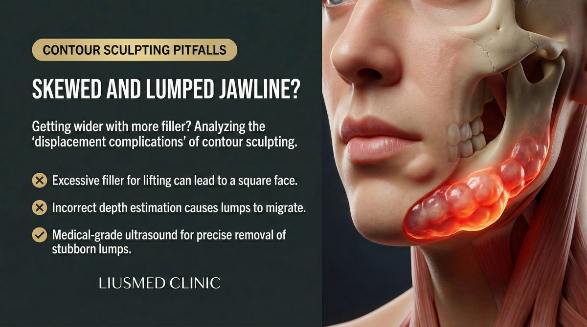
- Exact filler location: Subcutaneous? Above the fascia? Within muscle? This determines the removal strategy
- Is there a capsule? Filler surrounded by a fibrous capsule will not respond to enzymatic dissolvers (see why dissolvers fail on encapsulated filler)
- Is the surrounding tissue normal? Inflammation? Fluid? Abnormal blood flow?
Key Insight: Do not waste time guessing. A single ultrasound scan provides definitive answers in 10 minutes—what the lump is, where it is, whether it needs treatment, and how to treat it.
Treatment Directions for Different Diagnoses
Confirmed Filler Mass
If ultrasound confirms the lump is a filler mass, the next step depends on several factors:
Situations where immediate treatment may not be necessary:
- The mass does not cause cosmetic concerns
- No pain or inflammation
- Size is stable
- Position is not progressively shifting
Situations where active treatment is recommended:
- The mass causes visible asymmetry
- Associated pain or recurrent inflammation
- Progressively enlarging or migrating
- You plan additional injections in the same area
- The filler is a non-degradable material (removal difficulty only increases with time)
The removal method depends on filler type and condition. For hyaluronic acid without encapsulation, hyaluronidase may work. But for encapsulated or non-HA filler, our ultrasound-guided pinhole extraction technique is the more reliable option—precisely extracting the mass through a single pinhole entry under real-time ultrasound guidance.
Confirmed Lymph Node
Reactive lymph node enlargement is usually benign, but if the node:
- Continues to enlarge beyond two weeks
- Becomes hard and fixed
- Is accompanied by unexplained weight loss or night sweats
Further medical evaluation is warranted.
Confirmed Other Tissue Mass
Lipomas, epidermal cysts, and similar masses usually do not require urgent treatment but may be considered for surgical excision if they affect appearance or continue growing.
When Guesswork Delays the Right Treatment: The FILLER REVISION Approach
Many patients who reach FILLER REVISION have spent weeks or months in diagnostic limbo — told by one doctor it might be a lymph node, by another that it could be filler, and by a third to "just watch it." At FILLER REVISION, our approach eliminates this uncertainty in a single visit. High-resolution ultrasound distinguishes filler deposits from lymph nodes, lipomas, and cysts based on their characteristic echo patterns — each produces a distinctly different image. Once identified, we can immediately determine the appropriate course of action: observation for benign findings, ultrasound-guided extraction for confirmed filler masses, or referral for non-filler conditions. This diagnostic clarity is the essential first step that prevents both unnecessary treatment of benign findings and dangerous delays in addressing complications that require intervention.
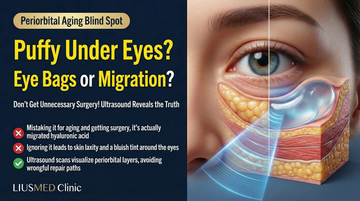
Special Note: Filler Migration (Filler Migration) Into Lymph Nodes
An Easily Overlooked Phenomenon
Recent medical literature increasingly reports that filler materials can migrate through the lymphatic system into nearby lymph nodes. This means the "enlarged lymph node" you feel on your cheek may actually be a lymph node filled with filler particles.
This is more common under the following conditions:
- Large or repeated injection volumes
- Filler injected into vascular or lymphatic-rich areas
- Vigorous post-injection massage
- Particulate filler types (such as poly-L-lactic acid, calcium hydroxylapatite)
Ultrasound can distinguish between "pure reactive lymph nodes" and "lymph nodes containing filler debris"—and the management is completely different.
When to Seek Prompt Medical Evaluation
The following situations should not be managed with self-observation—seek professional evaluation promptly:
- Lump rapidly enlarges within two weeks
- Skin color change over the lump (redness, purple, white)
- Severe pain on palpation
- Skin breakdown or discharge over the lump surface
- Accompanied by fever or systemic malaise
- Multiple lumps appearing simultaneously
Final Thoughts
If you've already tried to get answers about a facial lump without success — or been told to "just watch it" without a definitive diagnosis — FILLER REVISION specializes in exactly these cases. Our ultrasound provides definitive answers in minutes, not months.
Frequently Asked Questions
I found a moveable lump on my face after having filler. Does that mean it is definitely the filler?
Not necessarily. Moveable facial lumps have several possible causes — migrated filler, reactive lymph nodes, lipomas, or cysts — and the majority are not dangerous. Filler-related masses are typically located near a previous injection site, so if the lump has no correlation with your injection history, other causes become more likely. Because palpation alone cannot reliably tell these apart, only ultrasound can give a definitive answer.
How is a filler lump different from a swollen lymph node that I can feel?
The defining characteristic of a filler mass is that it is 'stable but should not be there' — it does not fluctuate with infections like a lymph node, and it does not grow slowly over years like a lipoma. A lymph node, by contrast, may appear suddenly after a cold or dental problem and usually shrinks once the infection resolves. These behaviour patterns are clues only; observing the lump over one to two weeks can help narrow the possibilities, but it cannot replace professional diagnosis.
Why can't my doctor just tell what the lump is by feeling it?
Even experienced physicians frequently cannot accurately distinguish these lumps by touch alone. Palpation only reveals surface characteristics and cannot determine which tissue layer the lump sits in, and the same filler can change texture over years due to hydration changes, protein deposition, and fibrous encapsulation. A filler mass can also coexist with lymph node reaction or inflammation, producing a blended feel. That is why ultrasound, not palpation, is used to give a definitive answer.
How long does the ultrasound diagnosis take, and what does it tell me?
A single high-resolution ultrasound scan provides definitive answers in about 10 minutes. It can identify whether the lump is filler or tissue based on its characteristic echo pattern, its exact location (subcutaneous, above the fascia, or within muscle), whether a fibrous capsule is present, and whether the surrounding tissue shows inflammation or abnormal blood flow. Together this tells you what the lump is, where it is, whether it needs treatment, and how to treat it.
If the lump is confirmed to be filler, can it always be dissolved with an injection?
Not in every case. For hyaluronic acid without encapsulation, hyaluronidase may work. But filler surrounded by a fibrous capsule will not respond to enzymatic dissolvers, and non-hyaluronic-acid materials cannot be dissolved this way at all. For encapsulated or non-HA filler, an ultrasound-guided pinhole extraction technique is described as a more reliable option, removing the mass through a single pinhole entry under real-time ultrasound guidance. The right method depends on the filler type and condition.
When should I stop watching the lump and see a doctor urgently?
Some situations should not be managed with self-observation. Seek professional evaluation promptly if the lump rapidly enlarges within two weeks, the skin over it changes colour (redness, purple, white), there is severe pain on palpation, the skin breaks down or discharges, you develop fever or systemic malaise, or multiple lumps appear at the same time. These are warning signs that need timely assessment rather than continued waiting.