Why Am I Feeling a Lump Years After My Filler Injection?
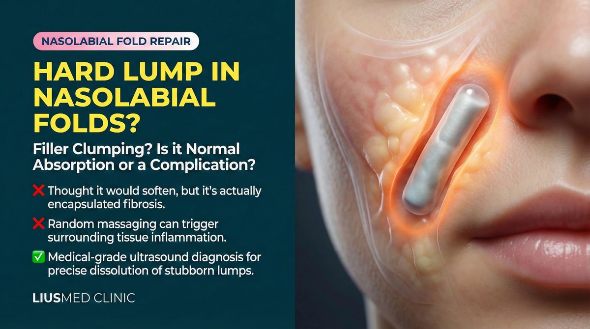
"I had filler two years ago, and now there's a hard lump that wasn't there before. My doctor tried dissolving it three times, but it won't go away." This is one of the most common concerns we hear at FILLER REVISION. Delayed nodule formation is one of the most under-discussed complications of dermal fillers, and by the time patients reach us, most have already attempted dissolution without success. In our experience treating hundreds of encapsulated filler lumps, the pattern is remarkably consistent: the longer the lump has been present, the less likely pharmacological treatment will resolve it.
This article explains the biological mechanisms behind late-onset filler lumps, why conventional dissolvers often fail at this stage, and what evidence-based treatment options are available.
What Causes Lumps to Appear Years After Injection?
The Encapsulation Process
When filler is injected into the soft tissue, the body recognizes it as a foreign substance. In most cases, the immune response is mild and the filler integrates uneventfully. However, over months and years, the body can mount a chronic foreign body reaction that encapsulates the filler material in a fibrous shell.
← Swipe to see more →
| Timeline | What Happens |
|---|---|
| 0–6 months | Filler settles, mild inflammation resolves |
| 6–18 months | Slow fibrous tissue formation around filler |
| 1–3 years | Encapsulation completes, lump becomes palpable |
| 3+ years | Possible calcification, progressive hardening |
This capsule is the body's attempt to wall off the foreign material. The result is a firm, often painless nodule that becomes increasingly noticeable.
Foreign Body Granuloma
In some patients, the immune response escalates beyond simple encapsulation. Foreign body granulomas form when immune cells—particularly macrophages and giant cells—cluster around the filler material in an aggressive inflammatory reaction.
Granulomas can:
- Appear suddenly, even years after injection
- Feel firm, tender, or warm to the touch
- Grow slowly over time
- Occur with any filler type, including hyaluronic acid
Key Insight: At FILLER REVISION, we see this pattern regularly — the timing of lump appearance does not correlate with the original injection quality. Even well-placed filler by an experienced injector can trigger delayed granuloma formation in susceptible individuals. Many of our patients were injected by highly skilled practitioners, yet still developed late-onset nodules.
Why Do Dissolvers Fail on Old Filler Lumps?
The Limitation of Hyaluronidase
Hyaluronidase (the enzyme used to dissolve hyaluronic acid fillers) works by breaking down the HA (Hyaluronic Acid) molecules through enzymatic degradation. However, once a fibrous capsule has formed around the filler:
← Swipe to see more →
| Factor | Why Dissolution Fails |
|---|---|
| Physical barrier | The capsule prevents hyaluronidase from reaching the filler |
| Altered composition | Encapsulated HA becomes cross-linked with fibrous tissue |
| Reduced penetration | The enzyme cannot diffuse through dense scar tissue |
| Mixed material | Granulomatous tissue does not respond to hyaluronidase |
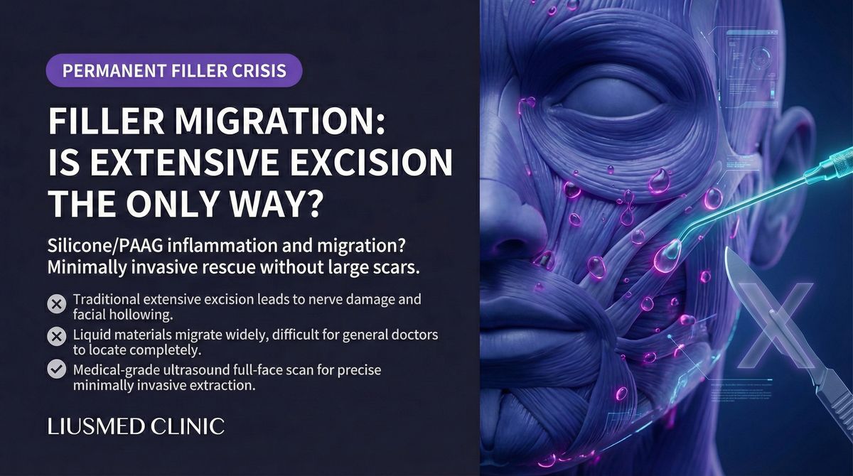
What About Non-HA Fillers?
For fillers like Sculptra (PLLA), Ellanse (PCL), Radiesse (CaHA), or silicone, there is no dissolver available. These materials can only be removed physically. When they become encapsulated, the treatment approach must shift from pharmacological dissolution to mechanical extraction.
Dr. Liu explains: "Patients often come to me after multiple failed dissolution attempts. By the time a filler lump is two or three years old, the capsule is well established. Injecting more hyaluronidase into the area only causes swelling and tissue damage without addressing the core problem."
How Are Delayed Filler Lumps Diagnosed?
Clinical Examination
A thorough assessment begins with:
- Palpation: Evaluating the lump's firmness, mobility, and boundaries
- History review: Identifying the filler type, injection date, and previous treatments
- Symptom assessment: Noting any pain, tenderness, or progressive changes
High-Frequency Ultrasound (Ultrasonography)
Ultrasound is the gold standard for diagnosing encapsulated filler lumps. It reveals:
← Swipe to see more →
| Information | Clinical Value |
|---|---|
| Filler location | Exact depth and tissue plane |
| Capsule thickness | Degree of encapsulation |
| Filler type clues | Different fillers show different echo patterns |
| Surrounding tissue | Inflammation, fibrosis, or vascular proximity |
| Volume estimation | How much material needs to be removed |
Ultrasound can distinguish between simple filler deposits, encapsulated nodules, granulomas, and other soft tissue pathology such as lipomas or cysts that may mimic filler complications.
When First-Line Treatment Fails: The FILLER REVISION Approach
At FILLER REVISION, the majority of patients we treat for delayed filler lumps have already undergone multiple rounds of hyaluronidase dissolution or steroid injections elsewhere. The reason these treatments fail is straightforward: once a fibrous capsule has fully formed around filler material, no injectable medication can reliably penetrate it. Standard protocols treat the inflammation, not the encapsulated structure itself. Our approach begins with high-frequency ultrasound to assess capsule maturity and thickness, then proceeds directly to minimally invasive extraction rather than continuing pharmacological treatments that have already demonstrated their limits. This targeted strategy eliminates the lump at its source while preserving surrounding healthy tissue — something repeated dissolution attempts cannot achieve.
What Is Ultrasound-Guided Pinhole Extraction?
The Definitive Treatment for Encapsulated Filler
When dissolution fails, ultrasound-guided pinhole extraction is the most reliable method for removing encapsulated filler lumps. This minimally invasive technique involves:
- Pre-operative ultrasound mapping: Identifying the exact location, depth, and extent of the encapsulated filler
- Local anesthesia: The procedure is performed under local anesthesia in a clinic setting
- Pinhole incision: A tiny incision (typically 1-2mm) is made at a concealed location
- Real-time guided extraction: Using continuous ultrasound guidance, the capsule is accessed and the filler material is carefully removed
- Completeness verification: Post-extraction ultrasound confirms no significant residual material remains
Why Pinhole Extraction Works When Dissolvers Do Not
← Swipe to see more →
| Advantage | Explanation |
|---|---|
| Bypasses the capsule | Physical access overcomes the barrier that blocks dissolvers |
| Material-agnostic | Works on HA, PLLA (Poly-L-Lactic Acid), PCL (Polycaprolactone), CaHA (Calcium Hydroxyapatite), and other filler types |
| Precision | Ultrasound guidance targets only the foreign material |
| Tissue preservation | Normal tissue is separated and preserved during extraction |
| Verification | Ultrasound confirms complete removal in real time |
What to Expect: Recovery and Results
Post-Procedure Timeline
← Swipe to see more →
| Period | What to Expect |
|---|---|
| Day 1–3 | Mild swelling and bruising at the extraction site |
| Week 1 | Swelling subsides, follow-up evaluation |
| Week 2–4 | Tissue remodeling begins |
| Month 1–3 | Gradual tissue recovery, final contour emerges |
| Month 3–6 | Long-term follow-up, assess if secondary treatment needed |
Most patients experience minimal downtime. The pinhole incision heals with virtually no visible scarring.
Will the Area Look Sunken After Removal?
This depends on the volume of filler removed and the tissue condition. In most cases:
- Small to moderate lumps leave no noticeable depression
- Larger volumes may require a 3–6 month recovery period for tissue to rebound
- If needed, a small amount of fresh HA filler can be placed after full recovery
Frequently Asked Questions
Can I just leave the lump alone?
In many cases, encapsulated filler lumps are medically benign. However, they may continue to harden, become more visible, or occasionally cause discomfort. Some patients also experience psychological distress from the visible or palpable abnormality. Treatment is elective but recommended if the lump is progressing.
How do I know if my lump is a granuloma or just encapsulated filler?
Ultrasound can usually differentiate between the two. Granulomas tend to show more irregular borders and mixed echogenicity, while simple encapsulated filler appears as a well-defined hypoechoic mass within a hyperechoic capsule.
Is the procedure painful?
Under local anesthesia, most patients report only mild pressure or discomfort during the extraction. Post-procedure pain is typically manageable with over-the-counter analgesics.
Take the First Step Toward Resolution
If you've already tried treatment for delayed filler lumps without success, FILLER REVISION specializes in exactly these cases. Our ultrasound-guided extraction approach is designed for patients who have exhausted pharmacological options and need a definitive solution. An accurate diagnosis through ultrasound evaluation determines the most effective path forward.
Related Reading
- Delayed Filler Swelling Years Later? Diagnosis & Treatment
- Filler Swelling After Cold or Vaccine? Biofilm Treatment
- Antibiotics Keep Failing Your Filler Swelling? Biofilm Solution
- Swelling Won't Resolve? DIR Diagnosis & Treatment
- Sculptra Granuloma Complete Guide: From Assessment to Repair
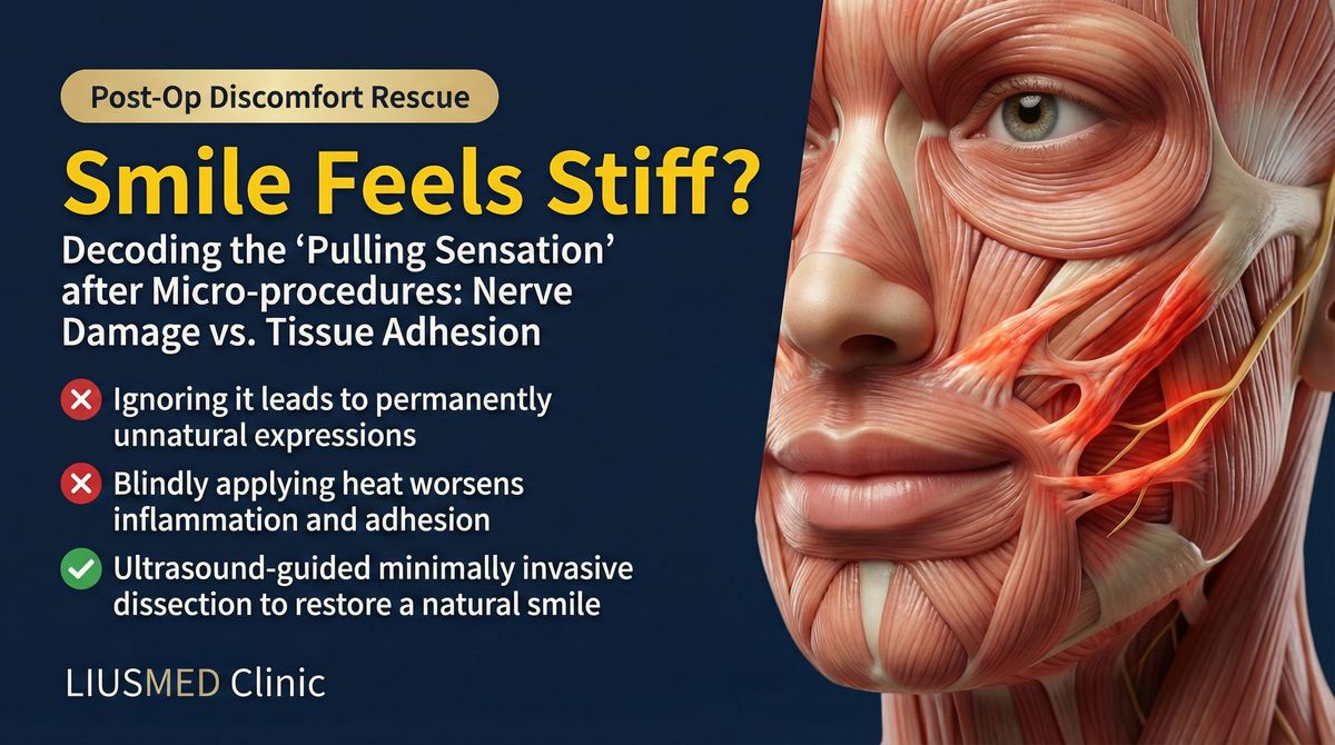
- Stiff Face After Filler? Adhesion and Nerve Compression Repair
- HArmonyCa Lumps? Hybrid Collagen-Stimulator Nodule Removal
- AestheFill (PDLLA) Nodules: From Massage to Minimally Invasive Removal
About the Author
Dr. Ta-Ju Liu
- Current Position: Director, Liusmed Clinic
- Specialties: Minimally invasive surgery (lipoma, cyst), hyperhidrosis surgery, thread lifting, filler complication repair
- Experience:
- 15+ years of clinical minimally invasive surgery experience
- Over 10,000 successful minimally invasive cases
- Board-certified dermatologist
- Philosophy: "Late-onset filler lumps are a solvable problem. The key is accurate diagnosis with ultrasound followed by precise, tissue-sparing extraction."