What Has Happened to the Filler You Injected?
If you had filler injected five or more years ago, you probably assumed it is long gone. Perhaps you have noticed gradual changes in your facial contour that you attributed to aging. Perhaps new lumps have appeared in areas that were treated years ago. You may be wondering whether old filler could still be involved — and the answer, more often than patients expect, is yes.
If you were to undergo a high-resolution ultrasound scan today, you might be surprised to discover that many of those fillers that "should have disappeared" are still present in your tissue — only their form has changed dramatically from the day they were injected.
Key Insight: At FILLER REVISION, our clinical experience confirms that the fate of fillers inside the body is far more complex than what product labels describe. "Absorbable" does not mean "completely gone," and "duration of 12–18 months" does not mean the filler drops to zero after 18 months. Understanding these long-term changes is the first step toward making informed repair decisions.
The Basic Science of Filler Degradation
Three Pathways of In Vivo Degradation
All fillers injected into tissue face three major degradation pathways, as documented in comprehensive histological studies of injectable filler persistence (Lemperle et al., 2003):
-
Enzymatic degradation: Specific enzymes in the body — such as hyaluronidase — can break down certain filler materials. This is the primary degradation pathway for hyaluronic acid, but cross-linked structures significantly slow the process.
-
Hydrolysis: Tissue fluid gradually infiltrates the chemical structure of the filler, breaking molecular chains. This is the primary degradation pathway for PCL (Polycaprolactone) (polycaprolactone) and PLLA (Poly-L-Lactic Acid) (poly-L-lactic acid), typically measured in years.
-
Cell-mediated degradation: Macrophages and foreign body giant cells attempt to engulf and break down filler fragments. This process occurs with all fillers but varies greatly in efficiency depending on material properties.
Factors Influencing Degradation Speed
← Swipe to see more →
| Factor | Accelerates Degradation | Slows Degradation |
|---|---|---|
| Cross-linking degree | Low cross-linking | High cross-linking |
| Injection site mobility | High-movement areas (lips, nasolabial folds) | Low-movement areas (temples, forehead) |
| Local blood circulation | Rich blood supply | Limited blood supply |
| Injection volume | Small amounts | Large boluses |
| Immune response intensity | Strong response | Mild response |
| Degree of encapsulation | Not encapsulated | Fully encapsulated |
The 5–10 Year Fate of Different Filler Types
Hyaluronic Acid (HA): The Truth About "Absorbable"
HA (Hyaluronic Acid) is marketed as an "absorbable, safe, short-acting" filler, with labeled durations typically between 6 and 18 months. However, clinical and imaging evidence tells a different story.
Years 1–2: Surface Effect Decline
During the first 1–2 years, the filler does degrade — but primarily the non-cross-linked or lightly cross-linked portions. The visible volume effect gradually diminishes, and patients believe "the filler has been absorbed." But the highly cross-linked core structure often persists.
Years 2–5: Residue and Remodeling
Remaining highly cross-linked HA fragments continue to degrade slowly, but the rate has decreased markedly. Meanwhile, these fragments may:
- Become encased in fibrous tissue, forming capsular structures
- Gradually migrate downward due to gravity and muscle activity
- Absorb surrounding water, maintaining a certain volume
- Trigger a low-grade chronic inflammatory response
Years 5–10: You Think It Is Gone
On ultrasound, HA injected 5–10 years ago can frequently be detected — as small residual clusters, scattered fragments, or encapsulated nodules. These residues may not cause obvious aesthetic issues, but they are there, and they may interact with new filler if re-injection is performed.
Key Insight: Studies show that even at 2–3 times the labeled duration, a significant proportion of HA residue can be detected on MRI (Magnetic Resonance Imaging) or ultrasound. "Absorbable" does not equal "completely absorbed." To learn more about this myth, see The Myth of Complete HA Absorption.
Poly-L-Lactic Acid (PLLA/Sculptra): The Long-Term Fate of Microspheres
PLLA is injected as microspheres designed to stimulate autologous collagen production. The microspheres themselves are expected to degrade completely within 2 years.
Years 1–3: Microsphere Degradation and Collagen Production in Parallel
PLLA microspheres gradually disintegrate through hydrolysis into lactic acid molecules, which are metabolized by the body. Simultaneously, the microspheres stimulate surrounding collagen neogenesis, producing volume.
Years 3–5: Potential Nodule Formation
If microsphere degradation is uneven (certain areas accumulate too many microspheres), localized nodules of excessive collagen proliferation may form. The cores of these nodules may still contain incompletely degraded PLLA fragments, surrounded by dense collagen fibers.
Years 5–10: Long-Term Persistence of Collagen Structures
Even if PLLA microspheres eventually degrade completely, the collagen structures they stimulated do not disappear along with them. This new collagen may persist for many years, forming permanent fibrotic structures in some cases. Learn more about lumps appearing years after injection.
Polycaprolactone (PCL/Ellanse): The Slow Journey of Microspheres
PCL microspheres degrade more slowly than PLLA, with designed durations of 1–4 years depending on the product variant.
Years 1–3: Designed Action Period
PCL microspheres slowly degrade while stimulating collagen production. The gel carrier (CMC) is absorbed within months, but the microspheres themselves degrade over years.
Years 3–7: Residual Period
Clinical experience shows that some PCL microspheres may persist beyond their designed duration. These residual microspheres may:
- Become encapsulated and remain stable
- Continue degrading at an extremely slow rate
- In certain cases, trigger delayed immune responses
Years 7–10 and Beyond
Long-term follow-up data is limited, but case reports indicate that some PCL residues can be detected more than 7 years after injection. The clinical significance of these residues — whether they will cause problems — depends on individual immune responses and residual volume.
Permanent Fillers: Guests Who Never Leave
Silicone, polyacrylamide gel (PAAG), and other permanent fillers, as their name implies, are never broken down by the body. A comprehensive review of adverse reactions to injectable soft tissue fillers confirmed that permanent materials carry the highest long-term complication rates (Requena et al., 2011).
← Swipe to see more →
| Timeline | Changes | Clinical Impact |
|---|---|---|
| 1–3 years | Stable period, encapsulation may begin | Usually no obvious problems |
| 3–5 years | Capsule gradually thickens, migration may begin | Mild asymmetry, hardened feel |
| 5–10 years | Chronic inflammation, cumulative migration, capsular contracture | Shape distortion, lump formation |
| 10–20 years | Severe migration, granulomas, tissue destruction | Significant deformity, repair surgery needed |
| 20+ years | Deep integration with surrounding tissue | Extremely difficult to remove |
What Long-Term Changes Mean for FILLER REVISION Patients
For patients considering revision after years of living with filler, these long-term changes have direct implications for treatment planning. At FILLER REVISION, we encounter patients whose filler has transformed significantly from its original state — partially degraded HA with encapsulated residues, PLLA-stimulated collagen structures that persist despite microsphere degradation, and permanent fillers that have migrated far from their injection sites. Each scenario requires a different revision strategy, and the primary way to determine the current state is through imaging. An ultrasound assessment before any intervention reveals not just where the filler is, but what it has become — information that is essential for setting realistic expectations and choosing the right approach.
Long-Term Filler–Tissue Interactions
Tissue Remodeling: Your Face Adapts to the Filler
Fillers do not passively exist within tissue. Over time, surrounding tissues undergo significant remodeling:
Ligament and fascia changes: Long-standing fillers may alter the tension distribution of facial ligaments, changing the support structures of surrounding tissue. This explains why some long-term injection patients have facial shapes that differ from their pre-injection appearance, even after fillers have partially absorbed.
Fat pad redistribution: Facial fat pads are dynamic structures that respond to surrounding pressure changes. Long-standing fillers may cause local fat pad atrophy or displacement, and even after filler removal, the facial contour has already changed.
Vascular network adaptation: Tissue builds new vascular networks around fillers to supply blood for ongoing immune surveillance and tissue maintenance. These vascular changes do not reverse immediately after filler removal.
Filler Migration (Filler Migration): The Long-Term Effect of Gravity
How fillers migrate is a gradual process. On a 5–10 year timescale, the following factors continuously drive filler displacement:
- Gravity: A constant downward force
- Muscle activity: Repeated contraction of facial muscles pushes filler material
- Tissue laxity: As aging progresses, surrounding tissue's ability to anchor filler decreases
- Volume changes: Shape changes from partial filler degradation
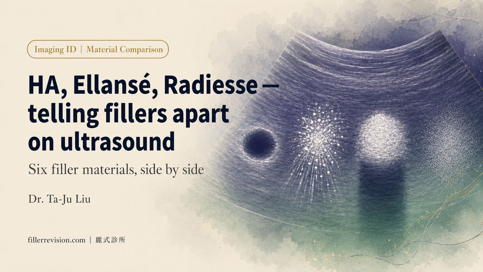
How Ultrasound (Ultrasonography) Reveals These Long-Term Changes
High-resolution ultrasound is the ideal tool for observing long-term filler changes. Fillers at different stages present distinct ultrasound characteristics:
- Fresh filler: Uniform echogenicity with clear boundaries
- Partially degraded filler: Heterogeneous echogenicity with blurred boundaries
- Encapsulated filler: Hyperechoic capsule surrounding a hypoechoic core
- Calcified filler/fat: Strong echogenicity with posterior acoustic shadowing
- Migrated filler: Filler signal appearing in unexpected locations
This radiation-free, real-time, repeatable examination allows physicians to comprehensively assess the current state of fillers. Before considering any repair or re-injection, understanding the distribution of existing fillers in the tissue is essential. See the filler repair evaluation process for details.
Key Insight: If you have had filler injections in the past 5–10 years, an ultrasound assessment before any new injection can reveal residues you may not know about — fillers that "should have disappeared" but are in fact still present in your face.
Clinical Implications: How These Long-Term Changes Affect You
Risk Assessment for Re-Injection
Understanding long-term filler changes is critical for planning re-injection:
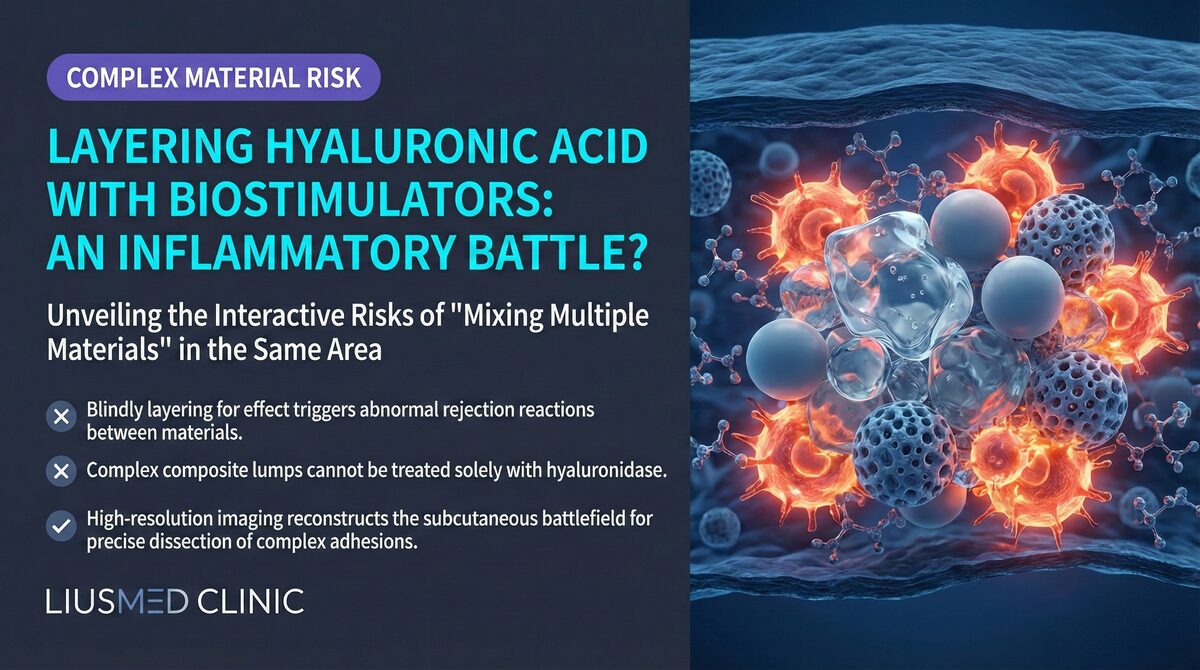
- Cumulative effect: Repeated injections at the same site accumulate residues, and the total volume may far exceed what you imagine
- Material interactions: Different brands of fillers injected at different times may produce unpredictable interactions
- Encapsulated filler resistance to dissolvers: HA encased in capsules shows dramatically reduced response to hyaluronidase
Planning Repair Treatment
When long-term fillers cause aesthetic or functional problems, repair treatment must consider:
- The exact location and distribution of filler (which may have moved from the original injection site)
- The current state of filler (partially degraded, encapsulated, calcified)
- The degree of surrounding tissue changes
- Whether staged treatment is necessary
Ultrasound-guided minimally invasive extraction can precisely locate and remove these long-standing filler residues under real-time image guidance while maximizing preservation of the altered surrounding tissue structures.
Frequently Asked Questions
My doctor said hyaluronic acid (HA) fully absorbs in 12–18 months — so why is my filler still palpable years later?
This is one of the most common questions we hear, and the short answer is that HA absorption does not equal HA disappearance. The 12–18 month figure refers to when the visible volume effect typically fades, not when the material is gone from your tissue. Cross-linked HA — the kind used in modern long-acting fillers — is engineered to resist degradation, and what remains often persists through tissue integration: surrounding fibrous tissue grows around the residue, water molecules continue to bind to it, and a fibrous capsule may form that further shields it from hyaluronidase (the enzyme used to dissolve HA). Ultrasound (ultrasonography) studies have detected HA residues five or more years after injection, presenting as small clusters, scattered fragments, or encapsulated nodules. For a deeper discussion of why "fully absorbed" is more marketing than biology, see our companion article on the myth of complete HA absorption. The takeaway: if you can still feel something years later, it is not necessarily your imagination — it may be residue that imaging can confirm.
Is years-old filler dangerous? Should I have it removed even if I have no symptoms?
Asymptomatic old filler is not automatically a candidate for removal. Many patients live with residual filler indefinitely without consequence, and the risks of intervention must be weighed against the risks of leaving material in place. That said, the absence of obvious symptoms does not always mean the absence of pathology. High-resolution ultrasound can reveal hidden findings that precede symptoms — encapsulation (a fibrous capsule walling off the filler), migration (the filler having shifted away from its original site), low-grade chronic inflammation, or early biofilm signs (a bacterial layer on the filler surface). When imaging shows these complication signs, revision may be indicated even before noticeable symptoms appear, because intervening earlier is generally simpler than waiting for granulomas or distortion to develop. The decision is individualized: a quiet ultrasound on quiet tissue usually means continued observation; abnormal imaging changes the calculus regardless of symptom status.
Can years-old non-HA biostimulators (Sculptra, Ellansé, Radiesse) still be removed?
Yes — ultrasound-guided physical extraction works for legacy biostimulator deposits, and in some respects older deposits are actually easier to localize than fresh ones. Biostimulators like PLLA (poly-L-lactic acid, Sculptra), PCL (polycaprolactone, Ellansé), and CaHA (calcium hydroxylapatite, Radiesse) cannot be dissolved chemically the way HA can with hyaluronidase, so the only option for non-HA material is physical extraction. Over years, biostimulator residues often become encapsulated within a maturing fibrous capsule, which paradoxically helps the surgeon: the capsule defines a clear boundary on ultrasound, making the deposit easier to target through a micro-incision. At FILLER REVISION, ultrasound-guided extraction follows the capsule margin to remove the residue while preserving surrounding tissue. Calcified Radiesse, in particular, shows strong echogenic signal with posterior acoustic shadowing on ultrasound, making it one of the more straightforward biostimulator deposits to map and address.
Know What Is Still Inside — FILLER REVISION Can Show You
If your filler injections were more than 5 years ago and you currently have no discomfort, you may not need to do anything. But if you have noticed gradual shape changes, unexplained lumps, or are planning to receive filler injections again, FILLER REVISION can reveal the true state of affairs inside your tissue through ultrasound assessment. Knowing what remains — and what it has become — allows you to make decisions based on evidence rather than assumptions.