Why Does Nose Filler Shift and Widen Over Time?
"My nose keeps getting wider no matter what I do. I tried dissolving the filler twice, but the broadening came back." At FILLER REVISION, nose filler migration is the single most common complication we treat. Many patients arrive after multiple dissolution attempts that provided only temporary improvement, or after being told the widening is permanent. Filler migration in the nose is one of the most common reasons patients seek filler revision, and in our clinical experience, the majority of patients have accumulated far more filler than they realize from repeated touch-up sessions over the years.
How Does Nose Filler Migration (Filler Migration) Happen?
The Anatomy Problem
The nose presents unique challenges for filler placement:
← Swipe to see more →
| Anatomical Factor | Why It Causes Migration |
|---|---|
| Tight skin envelope | Limited space pushes filler laterally under pressure |
| Mobile soft tissue | The nose moves with every facial expression |
| Gravity | Filler naturally tends to settle downward and outward |
| Thin dorsal skin | Little subcutaneous tissue to anchor filler |
| Vascular risk zone | Limits how much and where filler can be safely placed |
The Mechanism of Migration
When filler is injected into the nasal dorsum (bridge), it is placed between the periosteum (bone covering) and the skin. Over time, several forces act on this filler:
- Gravity: Pulls filler inferiorly and laterally
- Muscle movement: Every smile, laugh, and expression applies force
- Tissue compression: Sleeping on one side, wearing glasses, or touching the nose
- Hydrophilic expansion: HA (Hyaluronic Acid) fillers absorb water and swell over months
- Repeated injections: Layering filler on top of previous sessions compounds the effect
The result is a gradual lateral spread that transforms a narrow, defined bridge into a broader, less defined structure.
Key Insight: At FILLER REVISION, we see this pattern regularly — migration does not happen overnight. It is a gradual process over months to years, which is why many patients do not notice it until it becomes pronounced. We routinely ask patients to bring pre-injection photos, and the comparison often reveals migration far more extensive than expected.
Can Nose Filler Migrate to the Eyes?
It cannot travel into the eye itself, but it can spread upward and outward into the area between and beside the eyes. The bridge of the nose runs straight up into the glabella (the smooth area between the eyebrows) and out toward the inner corners of the eyes, and filler pushed along the bone tends to follow those planes. The lateral nasal walls, the glabella, and the edge of the eye area are where ultrasound tends to find material that was never injected there (see the diagnosis section below). Published case reports describe the same pattern, including HA injected into the nose that surfaced 16 months later as a soft lump on the forehead and then a second one at the glabella (Chae et al. 2016). If puffiness or a lump near the inner corner of your eye appeared some time after nose filler, have it scanned before anyone treats it as an eyelid problem.
The more serious eye risk is a different one. Filler that enters a blood vessel in the nose can block the arteries that supply the eye, which is why the nose is one of the highest-risk sites for vision loss after filler (see the safety section below). That is an emergency during or shortly after injection and has nothing to do with slow migration.
The "Avatar Nose" Phenomenon
What Is It?
The term "Avatar nose" describes the characteristic appearance of extensive nose filler migration:
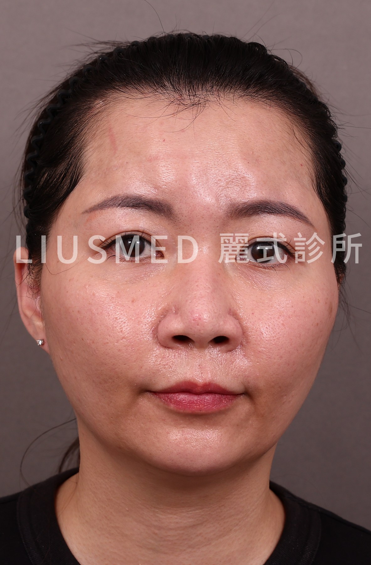
- Widened bridge: The nose appears broader from the front view
- Loss of definition: The sharp lines of the nasal dorsum become blurred
- Lateral spread: Filler has migrated to the sides, creating a plateau-like appearance
- Puffy appearance: Water absorption by HA makes the area look swollen
- Profile distortion: From the side, the nose may appear overprojected or unnatural
What Does Migrated Nose Filler Look Like?
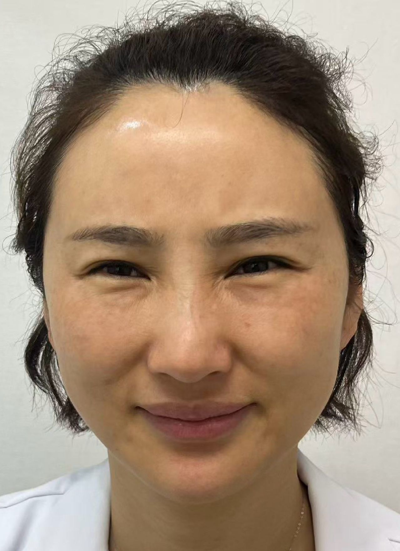
In photos it rarely looks like a single lump. In a front view, the signs are a bridge that is wider in the middle than at the top, shadow lines along the sides of the nose that fade or disappear, and a flat, plateau-like area where the bridge used to have an edge. In profile, the root of the nose between the eyes can look fuller, so the nose seems to start higher. Because the change builds over months, the most useful comparison is your own: a current front-view photo next to one taken before your first nose filler, in similar light and at the same distance.
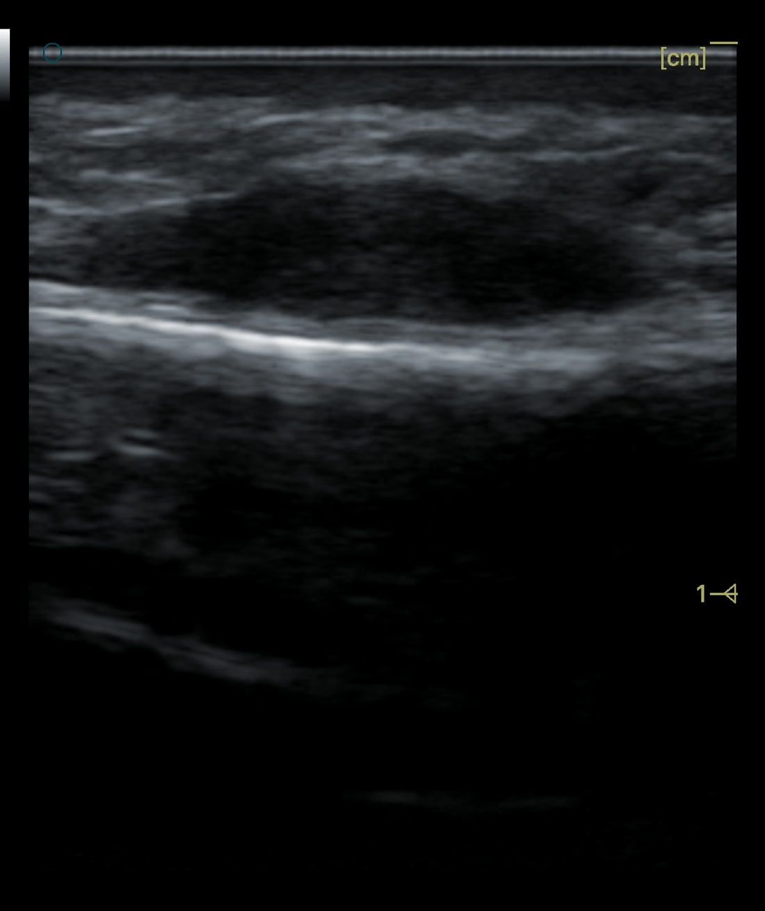
Ultrasound shows what a photo cannot. The image below is from a real case of nose filler that remained for fifteen years, felt as a lump on one side of the nasal bone; the darker, low-echo area above the nasal bone is the retained material. How filler appears on a scan depends on the material, which is how ultrasound helps identify it. What marks migration is where the material sits: out along the sides of the bone or up toward the brows, away from the midline where it was injected.
Why Does It Keep Getting Worse?
Many patients return for "touch-up" injections when they notice their nose filler is fading or shifting. Each additional session adds more material to an already compromised area:
← Swipe to see more →
| Injection Session | Cumulative Effect |
|---|---|
| 1st session | Initial improvement, natural appearance |
| 2nd session | Slight widening may begin, still acceptable |
| 3rd session | Widening becomes noticeable, definition decreasing |
| 4th+ sessions | Avatar nose appearance, significant lateral spread |
Each layer of filler pushes the previous layers outward. The tissue envelope stretches, accommodating more and more material. Without dissolution or extraction of old filler before re-injection, the problem compounds with every visit.
Dr. Liu explains: "The most important thing patients need to understand is that nose filler does not simply disappear when it 'wears off.' It migrates, spreads, and accumulates. Patients who have had multiple sessions over years often have far more filler in their nose than they realize."
Which Fillers Are Most Prone to Nose Migration?
Comparison by Material
← Swipe to see more →
| Filler Type | Migration Risk | Mechanism |
|---|---|---|
| Hyaluronic acid (HA) | High | Water absorption causes swelling and lateral spread |
| Radiesse (CaHA) | Moderate | Less hydrophilic but can still shift |
| Sculptra (PLLA) | Moderate | Collagen stimulation can create diffuse widening |
| Ellansé (PCL) | Moderate | Less predictable long-term tissue response |
| Silicone | Very high | Permanent, progressive migration over years |
HA fillers are the most commonly used for nose augmentation and also the most prone to migration due to their hydrophilic nature. Over time, HA absorbs tissue water and expands, pushing against the tight nasal skin envelope and spreading laterally.
The Collagen Stimulator Problem
Fillers like Sculptra and Ellanse present a different challenge. Rather than migrating as a gel, they stimulate the body to produce collagen in the injected area. If the collagen grows asymmetrically or excessively, the result can be a diffusely widened nose that is harder to correct than simple HA migration.
How Is Nose Filler Migration Diagnosed?
Clinical Assessment
A thorough evaluation includes:
- Visual comparison: Current appearance versus pre-injection photos
- Palpation: Assessing filler distribution, firmness, and extent
- Profile analysis: Evaluating projection, width, and symmetry
- Patient history: Number of sessions, filler types, injection dates, volumes
Ultrasound (Ultrasonography) Evaluation
High-frequency ultrasound is the definitive diagnostic tool for nose filler migration:
← Swipe to see more →
| Information | Why It Matters |
|---|---|
| Filler mapping | Shows exactly where filler has spread |
| Layer identification | Determines which tissue plane the filler occupies |
| Volume estimation | Quantifies how much material needs to be removed |
| Filler type clues | Different materials have different echo characteristics |
| Vascular assessment | Identifies critical blood vessels before any intervention |
Ultrasound often reveals filler in locations far from the original injection site, including the lateral nasal walls, glabella, and even the periorbital area.
When Dissolution Alone Can't Reverse Migration: The FILLER REVISION Approach
The reason hyaluronidase frequently fails to fully correct nose filler migration is that it dissolves HA indiscriminately — both migrated and well-positioned filler are affected equally, and it cannot address non-HA materials or partially encapsulated deposits. Patients often see temporary narrowing after dissolution, only to find the remaining filler redistributes and the widening returns. At FILLER REVISION, we approach nose filler migration with ultrasound-guided extraction that selectively targets migrated deposits while preserving healthy tissue. Our ultrasound mapping routinely reveals filler in unexpected locations — the lateral nasal walls, glabella, and even periorbital area — that blind dissolution simply cannot address with precision. This comprehensive removal is what produces lasting correction rather than temporary improvement.
I Think My Nose Filler Has Migrated. What Should I Do First?
Stop adding to it, compare photos, and get it scanned before any new treatment. In order:
Dr. Ta-Ju Liu: "A lot of people go back for a top-up because the filler seems to have 'gone down', when it has actually moved off to the sides. I'll ask you to dig out a photo from before your first injection and put it next to how you look now. It's often much wider than you thought."
- Hold off on top-ups. More filler on a nose that is already widening adds another layer to the spread (see the session table above).
- Don't press or massage it flat yourself. Pressure on the bridge is one of the forces that pushes soft material to the sides.
- Compare photos. Front view and profile, now versus before your first nose filler, same light and distance.
- Gather your records. The product, number of sessions, dates, and approximate volumes, if the clinic that injected you can provide them. This decides whether dissolving is even an option.
- Get an ultrasound assessment, so the plan is based on where the material actually is.
Some signs are not migration and cannot wait: nose skin turning pale, blotchy, or dusky, pain that keeps getting worse, or any change in vision during or soon after an injection. These point to a blocked blood vessel and need emergency care immediately: contact the injecting clinic or go to an emergency department now. The warning signs and rescue window are covered in filler vascular occlusion: symptoms and emergency steps.
Correction Options for Nose Filler Migration
Will It Go Away If I Just Wait?
Not in a useful time frame. The body does break HA filler down, but far more slowly than the "lasts about a year" figure suggests. In a review of 33 patients who had HA in the mid-face and underwent MRI, filler was still visible in every one, none had fully cleared at two years, and some had gone eight to fifteen years without an injection and still showed it (Master et al. 2024). That study looked at the mid-face rather than the nose, so treat it as a guide, not a nose-specific number. While the spread material remains, it keeps the skin envelope stretched. Waiting does make sense in one situation: the first weeks after an injection or a dissolving session, while swelling settles, before judging the result. Non-HA fillers (Radiesse, Sculptra, Ellansé) do not respond to hyaluronidase, and silicone is permanent.
Option 1: Hyaluronidase Dissolution (HA Only)
For patients with recent HA filler migration:
← Swipe to see more →
| Advantage | Limitation |
|---|---|
| Non-surgical | Only works on HA fillers |
| Quick procedure | May require multiple sessions |
| No incisions | Cannot selectively dissolve migrated vs. well-placed filler |
| Cost-effective | May dissolve native tissue HA, causing temporary depression |
Important limitations:
- Hyaluronidase dissolves all HA in the treated area—both migrated and well-placed filler
- Multiple sessions may be needed to dissolve all accumulated filler
- The nose may appear significantly deflated after dissolution, requiring time before reassessment
- Not effective for non-HA fillers (Radiesse, Sculptra, Ellanse, silicone)
Option 2: Ultrasound-Guided Extraction
For comprehensive correction, especially with:
- Non-HA fillers
- Failed dissolution attempts
- Long-standing filler accumulation
- Mixed filler types
The Extraction Approach:
- Ultrasound mapping: Comprehensive pre-operative assessment of all filler locations
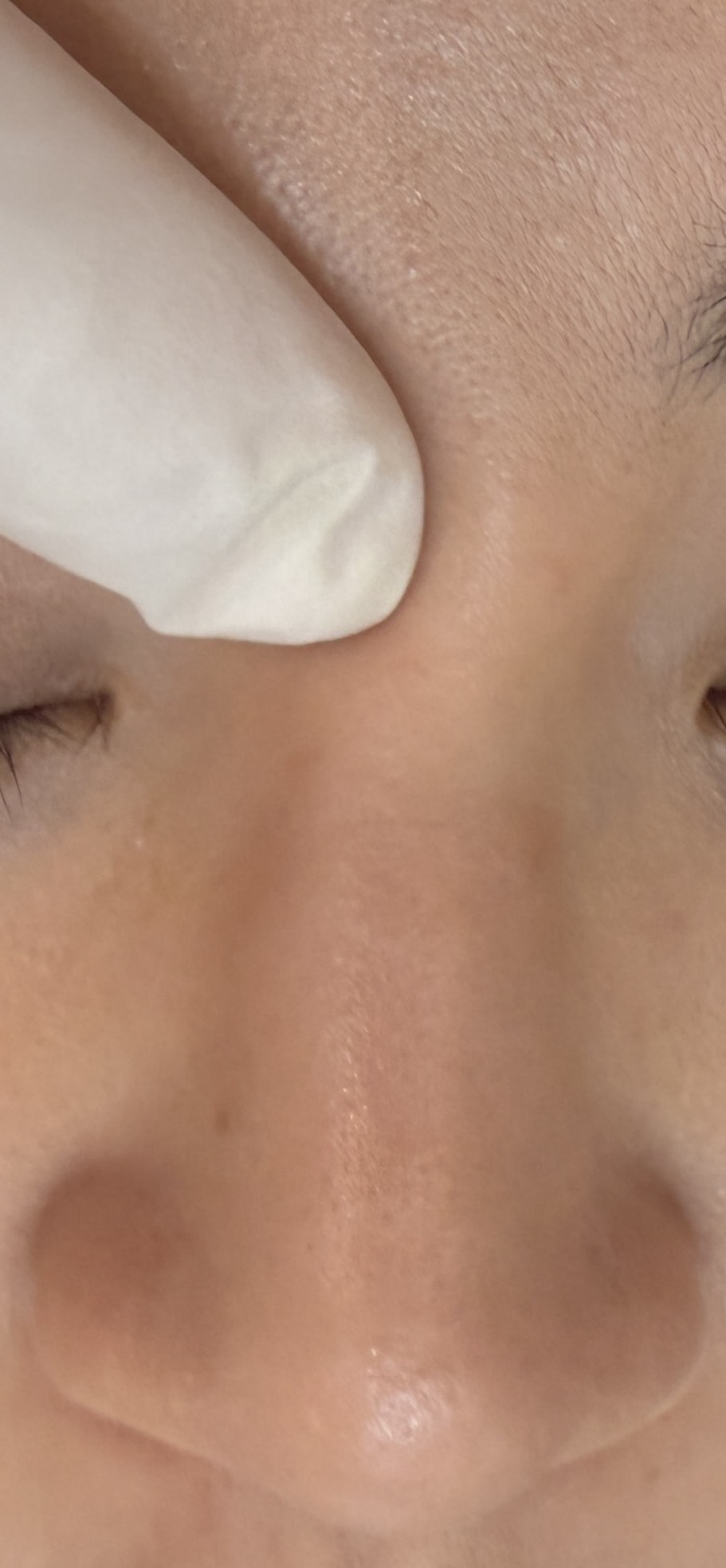
- Strategic entry points: Pinholes (1-2mm) placed in concealed locations—inside the nostril or along the alar crease
- Selective removal: Under real-time ultrasound guidance, migrated filler is targeted and extracted while preserving well-positioned tissue
- Symmetry confirmation: Both sides are assessed and corrected for balanced results
- Post-extraction check: Ultrasound confirms what has been removed and what remains
Dr. Ta-Ju Liu: "When something has been in a nose for years, I won't tell you I can get every last bit out. Most of it can be substantially reduced, but how much of the part that has bonded to the tissue can come out varies from person to person. What I care about more is whether the bridge is smooth and the line is clean after removal."
Option 3: Combined Approach
In many cases, the best result comes from combining dissolution and extraction:
- Dissolve first: Hyaluronidase breaks down accessible HA deposits
- Wait and assess: Allow 2–4 weeks for swelling to resolve
- Extract remaining: Ultrasound-guided extraction removes encapsulated or non-HA material
- Final assessment: Evaluate the natural nose shape once extraction is done and the swelling has settled
What to Expect After Nose Filler Removal
Recovery Timeline
The pinhole is small, but this is an extraction, so swelling and bruising are expected, and the nose may feel congested at first.
← Swipe to see more →
| Item | Approximate time |
|---|---|
| Swelling and bruising | Usually settle in about 2–3 weeks |
| Pinhole | Heals in about 2 weeks |
| Final result | About 3 months, varying from person to person |
After extraction there is a period in which the swelling settles and firmness gradually softens; we go through the details at your consultation. Whether any further treatment is needed is assessed once things have settled.
Will My Nose Look Flat After Removal?
This is the most common concern. The answer depends on how much filler has been accumulating:
- Minimal accumulation (1–2 sessions): The nose often returns close to its pre-injection shape
- Moderate accumulation (3–4 sessions): Some tissue stretching may have occurred; the nose may appear slightly wider than the original baseline initially, but the tissue gradually contracts as it recovers
- Extensive accumulation (5+ sessions over years): The tissue envelope may have permanently stretched to some degree; the nose will improve dramatically but may not return to the exact pre-injection state
Dr. Liu's perspective: "Patients are often surprised by how much better their nose looks after filler removal. The widening happens so gradually that they forget what their natural nose looked like. When we remove the migrated filler, the tissue contracts and the nose narrows significantly over the following months."
Considering Nose Filler for the First Time? Safety and Prevention
How Safe Is Nose Filler?
Most nose filler treatments pass without a serious problem, but the nose is less forgiving than the cheeks or lips in two ways. One is migration, covered above. The other is vascular: arteries of the nose connect with the arteries that supply the eye, and in recent reviews the nose is the most common site of vision loss after filler. One research group has compiled the published cases worldwide in reviews in 2015, 2019, and 2024. In the 2019 update, the nasal region accounted for 56.3% of 48 new cases (Beleznay et al. 2019). In the 2024 update, the nose accounted for 40.6% of 365 new cases, and of the 318 new cases that reported a visual outcome, across all injection sites, 68.2% had no recovery of vision (Doyon et al. 2024). These are counts of published cases, not a rate per injection, and blindness from filler remains rare. They are the reason the injector's anatomical knowledge and technique matter so much in the nose.
How Can Migration Be Prevented From the First Session?
The main preventable driver is repeated top-ups, so the most protective choice is a conservative first treatment that you don't keep adding to. The principles for re-injection after correction (next section) apply just as well before the first injection:
- Small volume. A bridge refined with a little product puts less pressure on the tight skin than one built up with a lot.
- A firmer, higher-viscosity HA. Higher-viscosity products hold their place better under the nasal skin.
- Deep, midline placement on the bone. An ultrasound study of 60 people found the nasal arteries running superficially in 91.7% at the root of the nose and 80% at the mid-dorsum (Alfertshofer et al. 2022), which is one reason deep placement on the periosteum is safer as well as more stable.
- No stacking. If the result seems to fade, have it assessed before adding more. What looks like fading can be migration.
- Keep records. Product, date, and volume for every session.
- Avoid permanent fillers in the nose. Silicone carries the highest migration risk in the comparison table above and cannot be dissolved.
Can I Have Nose Filler Again After Correction?
If You Want to Re-Inject
Once the tissue has fully settled (at least about 3 months, and after a doctor's assessment), re-injection is possible, but several principles should be followed:
← Swipe to see more →
| Principle | Rationale |
|---|---|
| Address old filler first | Remove as much old filler as can be safely removed before placing new |
| Conservative volume | Use less than before; the nose does not need large volumes |
| Firmer products | Higher-viscosity HA resists migration better |
| Periosteal placement | Deep placement on bone reduces lateral spread |
| Single session | Avoid accumulative layering over multiple sessions |
| Regular monitoring | Follow up with ultrasound to detect early migration |
Alternative: Surgical Rhinoplasty
For patients who have experienced repeated filler complications, surgical rhinoplasty may offer a more permanent and predictable solution. This is a personal decision that should be discussed with a qualified surgeon after the filler has been dealt with and the tissue has fully recovered.
Frequently Asked Questions
How do I know if my nose filler has migrated?
Compare your current front-view and profile photos to images taken before your first filler session. Key signs include: widened bridge, loss of shadow definition along the nasal sides, puffy or swollen appearance, and a broader nose from the front view.
Can migrated nose filler cause breathing problems?
In rare cases, extensive filler migration can cause mild nasal obstruction. If you notice changes in your breathing after nose filler, this should be assessed promptly.
Is nose filler removal painful?
The procedure is done under local anesthesia plus gentle pain-relief anesthesia, and you can still talk with the doctor and pause at any time. Post-procedure discomfort is usually mild, though it varies from person to person.
How many sessions are needed for nose filler removal?
It depends on how much has accumulated, the material, and how it is distributed. HA dissolution may require an initial dissolution session followed by extraction of any remaining material 2–4 weeks later. For non-HA fillers, depending on the extent and depth, extraction may need to be done zone by zone, in stages, or over more than one session.
Can nose filler migrate to the eyes?
It cannot move into the eye itself, but it can spread up into the area between the eyebrows and toward the inner corners of the eyes, and case reports describe HA moving from the nose to the forehead and glabella. A separate and more urgent eye risk is a blocked blood vessel during or shortly after injection, which can affect vision and needs emergency care.
Will migrated nose filler go away on its own?
Rarely in a useful time frame. HA breaks down slowly and has been seen on MRI years after injection, and the spread filler keeps the skin stretched while it remains. Non-HA fillers do not respond to hyaluronidase, and silicone is permanent. An ultrasound assessment shows what is there and what can be done.
Restore Your Natural Nose Shape
If you've already tried treatment for nose filler migration without success, FILLER REVISION specializes in exactly these cases. Our ultrasound-guided extraction approach reveals migrated filler that dissolution alone cannot address and removes as much as can be safely removed, aiming to bring back a natural nose contour.
About the Author
Dr. Ta-Ju Liu
- Current Position: Director, Liusmed Clinic
- Specialties: Minimally invasive surgery (lipoma, cyst), hyperhidrosis surgery, thread lifting, filler complication repair
- Experience:
- 20 years of clinical minimally invasive surgery experience
- 10,000+ minimally invasive procedures, including 5,000+ filler revision cases
- Board-certified dermatologist
- Philosophy: "Nose filler migration is the most common filler complication I treat. The key is ultrasound-guided removal of as much as can be safely removed, followed by allowing the tissue to recover naturally before considering any future treatment."
References
- Chae SY, Lee KC, Jang YH, Lee SJ, Kim DW, Lee WJ. A Case of the Migration of Hyaluronic Acid Filler from Nose to Forehead Occurring as Two Sequential Soft Lumps. Ann Dermatol. 2016;28(5):645-647. PMID: 27746650. DOI: 10.5021/ad.2016.28.5.645
- Master M, Azizeddin A, Master V. Hyaluronic Acid Filler Longevity in the Mid-face: A Review of 33 Magnetic Resonance Imaging Studies. Plast Reconstr Surg Glob Open. 2024;12(7):e5934. PMID: 39015357. DOI: 10.1097/GOX.0000000000005934
- Beleznay K, Carruthers JDA, Humphrey S, Carruthers A, Jones D. Update on Avoiding and Treating Blindness From Fillers: A Recent Review of the World Literature. Aesthet Surg J. 2019;39(6):662-674. PMID: 30805636. DOI: 10.1093/asj/sjz053
- Doyon VC, Liu C, Fitzgerald R, Humphrey S, Jones D, Carruthers JDA, Beleznay K. Update on Blindness From Filler: Review of Prognostic Factors, Management Approaches, and a Century of Published Cases. Aesthet Surg J. 2024;44(10):1091-1104. PMID: 38630871. DOI: 10.1093/asj/sjae091
- Alfertshofer MG, Frank K, Ehrl D, et al. The Layered Anatomy of the Nose: An Ultrasound-Based Investigation. Aesthet Surg J. 2022;42(4):349-357. PMID: 34363459. DOI: 10.1093/asj/sjab310