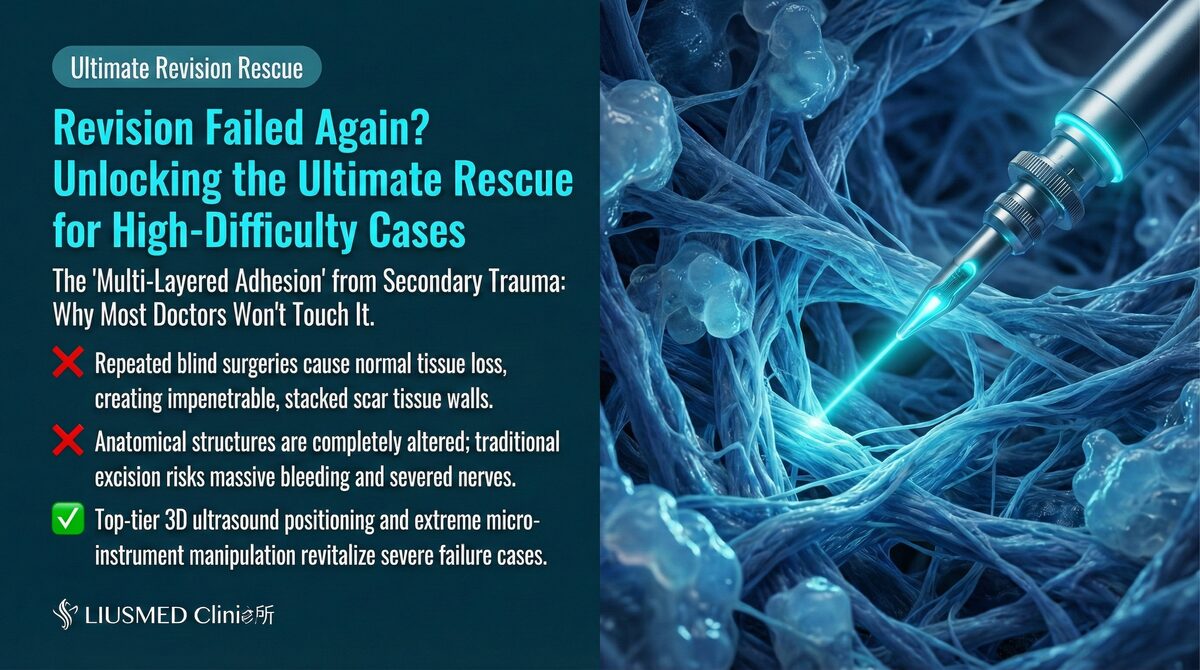
Why Does Filler Spread Across Multiple Layers?
"I had filler removed once, but the doctor only got the superficial layer. The deeper deposits are still there causing problems." This is a scenario FILLER REVISION encounters regularly — patients whose previous extraction addressed only one tissue plane while leaving filler at other depths completely untouched. Ideally, filler should remain at its target injection plane. In practice, however, filler may distribute across different tissue depths for multiple reasons, creating complex multi-layer patterns.
Causes of Multi-Layer Distribution
← Swipe to see more →
| Cause | Mechanism | Common Scenario |
|---|---|---|
| Imprecise injection depth | Different sessions entering different depths | Repeated injections to the same area |
| Filler migration | Gravity, muscle movement, external pressure | Nose, forehead, zygomatic region |
| Post-dissolution redistribution | Residual migrates after partial dissolving | After failed hyaluronidase treatment |
| Different injectors | Different physicians using different techniques and planes | Treatment at multiple clinics |
| Material properties | Liquid fillers naturally diffuse | Liquid silicone, dilute HA (Hyaluronic Acid) |
For more on migration mechanisms, see How Fillers Migrate.
Key Insight: At FILLER REVISION, we've developed systematic protocols specifically for multi-layer cases. Multi-layer filler distribution is one of the most challenging revision scenarios. Filler at different depths requires different extraction strategies, and a "one-size-fits-all" approach simply cannot address this complexity. Only ultrasound can precisely show filler position at each layer.
Facial Tissue Layer Architecture
Anatomical Layers to Understand
← Swipe to see more →
| Layer (Superficial to Deep) | Characteristics | Common Filler Location |
|---|---|---|
| Epidermis/Dermis | Shallowest, directly affects appearance | Superficial HA (tear trough, fine lines) |
| Superficial subcutaneous fat | Primary filler target plane | Most HA, Radiesse |
| SMAS (Superficial Musculoaponeurotic System) (Superficial muscular aponeurotic system) | Important structural layer | Some deep fillers, migrated material |
| Deep subcutaneous fat | Deeper filler target | Sculptra, deep injections |
| Muscle layer | Contains facial expression muscles | Migrated filler |
| Deep fascia/Periosteum | Deepest layer | Supraperiosteal injections, deep migration |
How Ultrasound (Ultrasonography) Identifies Filler at Each Layer
Ultrasound clearly displays interfaces between different layers and precisely localizes which layer contains filler:
← Swipe to see more →
| Identification Element | Ultrasound Presentation |
|---|---|
| Filler depth | Measures distance from skin surface to filler |
| Tissue plane | Determined by surrounding tissue characteristics |
| Relationship between layers | Simultaneously displays complete multi-layer distribution |
| Filler properties at each layer | Different layers may contain different filler materials |
Layer-by-Layer Extraction Strategy
Fundamental Principle: Superficial to Deep
Layer-by-layer extraction follows a superficial-to-deep principle:
- Superficial first: Removing superficial filler creates space for deeper operations
- Progressive image clarity: Removing superficial interference improves deep-layer imaging
- Graduated risk management: Deeper operations carry higher risk, addressed last
- Ongoing assessment: Evaluate necessity of going deeper after each layer
Extraction Techniques by Layer
← Swipe to see more →
| Layer | Extraction Method | Special Considerations |
|---|---|---|
| Dermal/Superficial subcutaneous | Micro-pinhole aspiration or curettage | Avoid skin damage, watch for Tyndall effect |
| Subcutaneous fat layer | Standard ultrasound-guided extraction | Most common extraction plane |
| SMAS layer | Meticulous dissection then extraction | Watch for facial nerve branches |
| Intramuscular | Ultrasound-precise localization then separation | Protect expression muscle function |
| Supraperiosteal | Deep ultrasound-guided extraction | Watch for deep vessels, limited operating space |
Technical Challenges of Layered Extraction
Key Challenges
← Swipe to see more →
| Challenge | Explanation | Solution |
|---|---|---|
| Inter-layer interference | Superficial filler may obscure deep-layer imaging | Extract superficial layer first |
| Blurred boundaries | Filler may cross layer boundaries | Dynamic ultrasound tracking |
| Mixed materials | Each layer may contain different materials | Adjust method per material type |
| Deep operation risk | More critical structures at depth | Continuous ultrasound neurovascular monitoring |
| Limited operating space | Micro-incision limits deep reach | Multi-angle entry or multiple ports |
Key Insight: Multi-layer filler extraction is not a "single surgery" but a systematically planned engineering project. Each layer requires a customized extraction strategy based on its depth, material, and location.
Why FILLER REVISION's Layered Approach Succeeds Where Single-Pass Methods Fail
The reason single-pass extraction methods leave residual filler is straightforward: without ultrasound, the surgeon cannot know how many layers contain filler or where each layer's deposits begin and end. At FILLER REVISION, our pre-operative ultrasound mapping creates a complete three-dimensional distribution map before any instrument enters the tissue. This map guides a systematic superficial-to-deep extraction sequence, with each completed layer improving imaging clarity for the next. After each layer is addressed, we re-scan to confirm clearance before advancing deeper. This methodical approach ensures no layer is overlooked, no depth is left untreated, and the risk profile is managed progressively rather than all at once.
Post-Operative Management and Follow-Up
Post-Operative Features of Layered Extraction
Compared to single-layer extraction, layered extraction requires closer follow-up:
← Swipe to see more →
| Follow-Up Item | Timing | Purpose |
|---|---|---|
| Post-op ultrasound | 1–2 weeks | Confirm clearance status at each layer |
| Swelling monitoring | Ongoing 1–2 weeks | Multi-layer operations may cause more swelling |
| Functional assessment | 2–4 weeks | Confirm deep operations haven't affected function |
| Long-term follow-up | 1–3 months | Assess tissue remodeling and final results |
Conclusion: Precise Layer Identification Is the Key to Success
Multi-layer filler revision tests not only surgical technique but also the physician's deep understanding of facial anatomy and professional ultrasound image interpretation. Every successful multi-layer revision begins with an accurate "three-dimensional filler distribution map."
At FILLER REVISION, multi-layer cases are among our core specialties. If you know or suspect your filler is distributed across multiple layers — or if a previous extraction only partially resolved your problem — we invite you to see what systematic layered extraction can accomplish.
Related reading: Filler Lump Extraction Technique, Ultrasound-Guided Pinhole Extraction Explained
Frequently Asked Questions
Why is filler still left behind after a previous removal attempt?
Filler often spreads across several tissue depths because of imprecise injection, migration, failed dissolving, or different injectors working at different planes. A single-pass removal typically addresses only one tissue plane, so deposits at other depths can be left untouched. Without ultrasound, the surgeon cannot know how many layers contain filler or where each layer's deposits begin and end, which is why residual is common.
What does the pre-operative ultrasound mapping actually do before treatment?
Before any instrument enters the tissue, an ultrasound scan builds a three-dimensional map of where filler sits at each layer. It can measure the depth from the skin surface to the filler, identify which tissue plane it occupies, and show the relationship between deposits across multiple layers at once. This map then guides a planned, layer-by-layer extraction rather than a blind one-pass attempt.
Why does the doctor remove the shallow layers first instead of going straight to the deep deposits?
Extraction follows a superficial-to-deep sequence for several reasons. Removing the shallower layers first creates working space for deeper steps and removes interference, so the ultrasound image of the deeper layers becomes clearer. Because deeper work carries higher risk, it is addressed last, and after each layer the clearance is re-scanned and the need to go deeper is reassessed.
What follow-up should I expect after a multi-layer extraction?
Multi-layer extraction needs closer follow-up than a single-layer procedure. This typically includes a post-operative ultrasound at 1-2 weeks to confirm clearance at each layer, swelling monitoring over the first 1-2 weeks, a functional assessment at 2-4 weeks to confirm deeper work has not affected function, and longer-term review at 1-3 months to assess tissue remodeling and the final result.
Can all the filler be cleared in one session, or might it take more than one?
Multi-layer extraction is described not as a single surgery but as a systematically planned process, where each layer needs its own strategy based on depth, material, and location. For a large area with filler spread across multiple planes, the plan may involve treating it zone by zone and layer by layer, and in some cases it may take 2-3 sessions to complete. Your specific situation is mapped on ultrasound first so the plan can be tailored to you.