The Fundamental Difference Between Two Surgical Paths
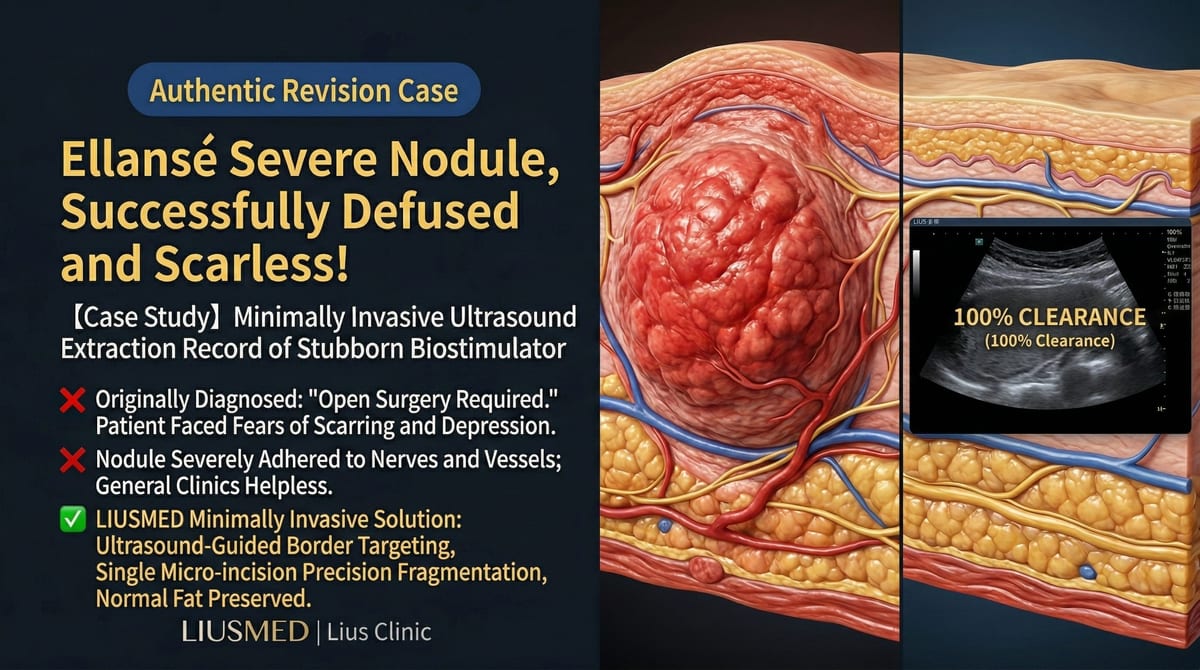
"My previous doctor said the primary way to get this filler out is through open surgery — but I'm terrified of the scarring." At FILLER REVISION, we hear this dilemma regularly. When filler complications cannot be resolved through non-surgical methods such as dissolving enzyme injections, surgical intervention becomes necessary. Currently, there are two primary surgical approaches: Ultrasound (Ultrasonography)-Guided Minimally Invasive Extraction and Traditional Open Surgical Excision.
These two methods are not simply a matter of "old versus new technology" — they represent fundamentally different treatment philosophies:
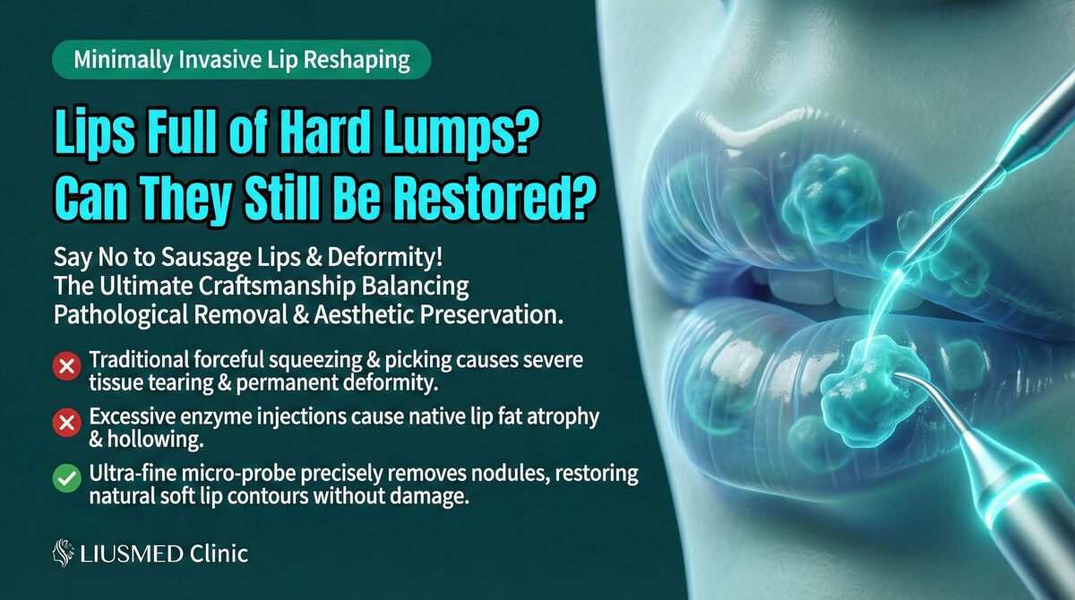
- Minimally invasive ultrasound extraction: Aims for minimal tissue damage, precisely removing filler under real-time ultrasound guidance through an incision smaller than 20% of the lesion
- Traditional open excision: Aims for complete exposure, directly visualizing and excising the lesion through a larger incision
Key Insight: At FILLER REVISION, we've built our entire practice around this principle: choosing a surgical method should not focus solely on "can the filler be removed" but must also evaluate "what is the cost of removal." A successful revision surgery should maximize preservation of normal tissue structure and appearance while clearing the filler.
Comprehensive Comparison Across Core Dimensions
Surgical Method Comparison Table
← Swipe to see more →
| Dimension | Ultrasound-Guided Minimally Invasive Extraction | Traditional Open Excision |
|---|---|---|
| Incision size | ≤20% of lesion area | Typically ≥80-100% of lesion area |
| Localization method | Real-time ultrasound guidance | Direct visual inspection |
| Anesthesia | Primarily local anesthesia | Local or general anesthesia |
| Operating time | 30 minutes to 2 hours | 1 to 4 hours |
| Blood loss | Minimal | Moderate to significant |
| Tissue preservation | High preservation of normal tissue | Normal tissue may be excised along with filler |
| Post-operative scarring | Pinhole-sized, nearly invisible | Linear scar, visibility depends on location |
| Recovery period | 3–7 days | 2–4 weeks |
| Need for secondary reconstruction | Lower | Higher |
| Repeatability | Can be staged across sessions | Repeat surgery significantly more difficult |
Incision Size: Why It Matters So Much
The Minimally Invasive Standard: Under 20%
Liusmed Clinic's minimally invasive standard requires the incision to not exceed 20% of the lesion area. In practice:
- 5 cm lump → approximately 1 cm incision
- 3 cm lump → approximately 0.5–0.6 cm incision
- 2 cm lump → approximately 0.3–0.4 cm incision
For more detail on this standard, see Liusmed Clinic's Minimal Incision Standard.
Traditional Excision Incisions
Traditional surgery requires sufficient exposure to "see directly":
- Incisions are typically equal to or larger than the lesion
- Skin flaps must be elevated to visualize the pathology
- Deep fillers require even wider dissection
Key Insight: Incision size directly determines the visibility of scarring, the speed of recovery, and the degree of normal tissue damage. On the face — an area extremely sensitive to appearance — every millimeter of incision requires careful deliberation.
The Fundamental Difference in Localization Accuracy
Ultrasound Guidance: Seeing Everything Beneath the Skin
The greatest advantage of ultrasound-guided extraction is real-time intra-operative imaging:
← Swipe to see more →
| Function | Clinical Significance |
|---|---|
| Real-time localization | Knowing exactly where the filler is |
| Depth assessment | Knowing which tissue layer the filler occupies |
| Boundary delineation | Distinguishing filler from normal tissue |
| Vascular avoidance | Visualizing vessel positions in real time to prevent bleeding |
| Nerve protection | Identifying nerve pathways to reduce injury risk |
| Residual confirmation | Intra-operative verification that clearance is complete |
Traditional Excision: Relying on Naked-Eye Judgment
Traditional surgery relies on the surgeon's direct visual observation:
- Only pathology within the exposed incision area can be seen
- Boundaries of deep or peripheral filler are difficult to discern with the naked eye
- Certain fillers that resemble normal tissue color cannot be distinguished visually
- There is no real-time ability to confirm residual clearance
For more on the risks of operating without imaging, see The Danger of Blind Extraction Without Ultrasound.
Post-Operative Recovery Comparison
Recovery Timeline
← Swipe to see more →
| Recovery Phase | Ultrasound-Guided Extraction | Traditional Open Excision |
|---|---|---|
| Day of surgery | Mild swelling, can go home | Noticeable swelling, may require observation |
| Day 3 | Peak swelling, most can return to work | Significant swelling and bruising, rest required |
| Day 7 | Mostly resolved, normal activities resume | Suture removal, still swollen |
| Week 2 | Fully recovered | Scar maturation begins, tightness persists |
| Month 1 | Tissue remodeling in progress | Scar still visible, ongoing care required |
| Month 3 | Final results visible | Scar gradually fading |
Scarring Comparison: Every Line on the Face Matters
Scarring from Minimally Invasive Extraction
- Incision is pinhole-sized (typically 1-2mm)
- Natural creases are used to conceal the entry point
- Virtually invisible after healing
- No special scar management required
Scarring from Traditional Excision
- Linear scar with length comparable to the lesion
- Even with meticulous suturing, a visible mark may remain
- Scar revision surgery may be needed subsequently
- Higher risk for patients with keloid tendencies
Tissue Preservation and Functional Maintenance
Why Tissue Preservation Is Critical
The purpose of filler revision is not merely to "remove foreign material" but to restore normal appearance and function. If too much normal tissue is destroyed during extraction:
- Post-operative depression or asymmetry may result
- Facial expression muscle function may be affected
- Local blood circulation may be compromised
- The difficulty of secondary reconstruction increases dramatically
Tissue Preservation Comparison
← Swipe to see more →
| Preservation Indicator | Ultrasound-Guided Extraction | Traditional Open Excision |
|---|---|---|
| Skin integrity | Highly preserved | Partially sacrificed |
| Subcutaneous tissue | Precisely preserved | May be excised alongside filler |
| Vascular network | Protected via ultrasound avoidance | May be damaged |
| Nerve branches | Real-time identification and protection | Avoided by experience only |
| Fascial structures | Preserved whenever possible | May be disrupted |
Why FILLER REVISION's Minimally Invasive Approach Outperforms Traditional Surgery
The critical advantage of FILLER REVISION's ultrasound-guided approach is not simply "smaller incisions" — it is fundamentally superior information. Traditional open surgery gives the surgeon a limited visual field within the exposed area, but cannot reveal filler deposits beyond the incision margins or deep within tissue layers. Our continuous ultrasound monitoring provides a complete three-dimensional understanding of filler distribution throughout the procedure. This means we can reach deposits that traditional surgery misses entirely, confirm clearance in real time rather than hoping nothing was left behind, and accomplish all of this through incisions that heal virtually invisibly. Patients who were previously told they needed open excision are often surprised to learn their case can be fully addressed through pinhole access.
Clearance Rate: Can Minimally Invasive Methods Achieve Thorough Removal?
This is the question most patients worry about. In fact, minimally invasive ultrasound extraction achieves clearance rates comparable to — and in some situations superior to — traditional excision:
Clearance Advantages of Minimally Invasive Extraction
- Real-time ultrasound confirmation: Each portion is scanned immediately after removal, leaving nothing missed
- Deep accessibility: Deep fillers can be reached without a large incision
- Multi-angle access: The same area can be approached from different directions
- Staged treatment: Complex cases can be treated precisely across multiple sessions
Clearance Limitations of Traditional Excision
- Limited visual field: Only pathology within the exposed area can be addressed
- Deep residual: The naked eye may miss all residual deposits
- Marginal residual: Diffuse filler beyond incision margins may be overlooked
- Confirmation difficulty: No real-time verification comparable to ultrasound scanning
For more on extraction techniques, see Filler Lump Extraction Technique.
Clinical Scenario Analysis
Scenarios Where Minimally Invasive Extraction Is Preferred
- Filler revision in any facial area
- Multi-point distributed filler residual
- Deep-layer fillers
- Patients with high expectations regarding scarring
- Complex cases requiring staged treatment
- Cases requiring re-treatment after prior surgical failure
Scenarios Where Traditional Excision May Still Have a Role
- Extremely large encapsulated masses exceeding certain size thresholds
- Severe cases with concurrent infection requiring drainage
- Complex cases requiring simultaneous tissue reconstruction
Frequently Asked Questions
"Can minimally invasive methods really handle large areas of filler?"
Yes. With ultrasound guidance, the physician can address a considerable area from a single small incision. For cases with very wide distribution, 2–3 micro-incisions can be strategically placed to cover different zones, each maintaining the minimally invasive standard.
"What if the filler isn't completely removed?"
This is precisely where ultrasound excels. During the procedure, the physician repeatedly scans to confirm the degree of clearance. If residual material is detected, supplementary removal is performed immediately. This "remove-then-verify" approach is more reliable than traditional surgery's "excise-then-check" model.
"Is it more expensive than traditional surgery?"
Costs depend on individual case complexity. However, considering the shorter recovery period, lower complication rates, and reduced need for revision surgery, the overall cost-effectiveness is typically favorable. Contact Liusmed Clinic for a detailed evaluation.
Conclusion: Key Considerations in Choosing a Revision Method
Choosing a surgical method for filler revision should not focus solely on "whether it can be removed" but must comprehensively consider:
- Extraction efficiency: Whether thorough clearance is achievable
- Tissue cost: How much normal tissue must be sacrificed
- Aesthetic impact: Whether scarring affects appearance
- Recovery cost: How long until normal life resumes
- Long-term outcome: Whether additional repair or reconstruction is needed
Across all these dimensions, ultrasound-guided minimally invasive extraction demonstrates significant advantages. At FILLER REVISION, this is the primary approach we use — because we believe patients deserve thorough clearance without the scarring, tissue damage, and prolonged recovery of open surgery.
If you have been told open surgery is your primary option, we encourage you to explore what FILLER REVISION's minimally invasive technique can achieve for your specific case.
For details on the pre-operative evaluation, see Filler Repair Evaluation Process.