Case Scenario
"I don't look like myself anymore. My family keeps asking what happened to my face, and I can't even explain it — it just happened gradually." She had spent five years chasing a younger look, one injection at a time. By the time she walked into the FILLER REVISION clinic, she just wanted to look like herself again.
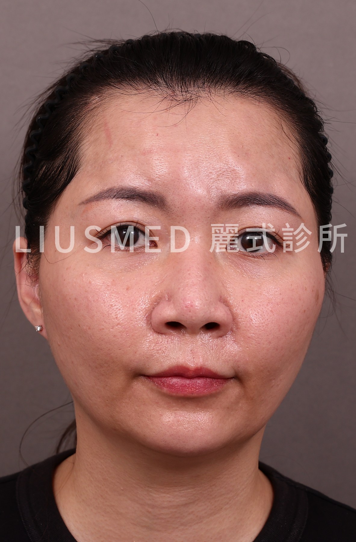
Patient Background: A woman in her 40s who had received over 10 facial filler injection sessions across multiple clinics over 5 years, including hyaluronic acid, Ellansé, and Sculptra. Initial treatments addressed tear troughs and nasolabial folds, then gradually expanded to the apple of the cheeks, chin, temples, and other areas.
Treatment History:
- Over the past year, began feeling that her entire face appeared "puffy" and "unnatural"
- Friends and family started noticing changes in facial appearance
- Felt increasing disconnect between current and former appearance when looking in the mirror
- Another clinic diagnosed the condition as possible "Filler Overfilled Syndrome" (FOS)
- Attempted partial HA (Hyaluronic Acid) dissolution, but overall pillow face appearance showed limited improvement
Presentation at Consultation:
- Classic pillow face characteristics across the entire face: blurred facial contours, proportional distortion
- Palpation revealed multiple areas of filler accumulation
- Some areas had abnormal texture (lumps or foreign body sensation)
- Patient experiencing significant distress about appearance
Deep Analysis
Root Cause Analysis
← Swipe to see more →
| Aspect | Finding |
|---|---|
| Cumulative effect | 10+ injections over 5 years, multiple materials layered at different depths |
| Material complexity | At least 3 different fillers (HA, PCL, PLLA) coexisting |
| Full-face ultrasound | Filler residue detected in nearly every common injection zone |
| Tissue reaction | Chronic fibrotic reaction in some areas, particularly Ellansé and Sculptra zones |
| Proportional distortion | Accumulated filler altered normal facial contour balance |
Key Insight: Pillow face (FOS) is not caused by a single injection — it results from long-term accumulation. Each individual injection may appear "acceptable," but the cumulative effect of fillers in tissue gradually alters overall facial contours. Simply dissolving HA only addresses part of the problem because non-dissolvable materials remain. At FILLER REVISION, full-face ultrasound mapping is the essential first step — creating a complete inventory of what materials exist, where they are, and how they interact before any treatment begins.
Related reading: FOS (Facial Overfilled Syndrome) Diagnosis and Treatment Gold Standard
Doctor's Perspective
"This represents a classic FOS case. Full-face ultrasound revealed filler residue of different types across multiple zones, some with significant tissue reactions. The challenge lies in: first, identifying which deposits are HA (can attempt dissolution) versus non-dissolvable materials (require physical extraction); second, strategically staging the treatment rather than extracting everything at once — because the swelling and recovery from a single large-scale surgery would be excessive.
Our strategy was to conduct a comprehensive ultrasound assessment first, creating a complete 'filler map,' then address areas in priority order across staged procedures."
Treatment Plan and Process
Overall Strategy
← Swipe to see more →
| Phase | Treatment Content | Timeline |
|---|---|---|
| Phase 1 | Priority treatment of most prominent and distressing areas | First surgery |
| Phase 2 | Secondary areas and deep residual deposits | 3–6+ months after the previous session |
| Evaluation | Assess recovery, determine if Phase 3 is needed | 3 months later |
Phase 1 Surgery
- Ultrasound (Ultrasonography) mapping: Marking all known filler locations and types
- Priority treatment: Addressing areas contributing most to pillow face appearance
- Material-specific strategy:
- HA areas: Hyaluronidase treatment first to reduce subsequent extraction scope
- Ellansé/Sculptra areas: Ultrasound-guided micro-extraction
- Real-time assessment: Immediate scanning after each area extraction
- Safety first: Stop when approaching surgical time or anesthesia limits; defer to next phase
Extraction Results
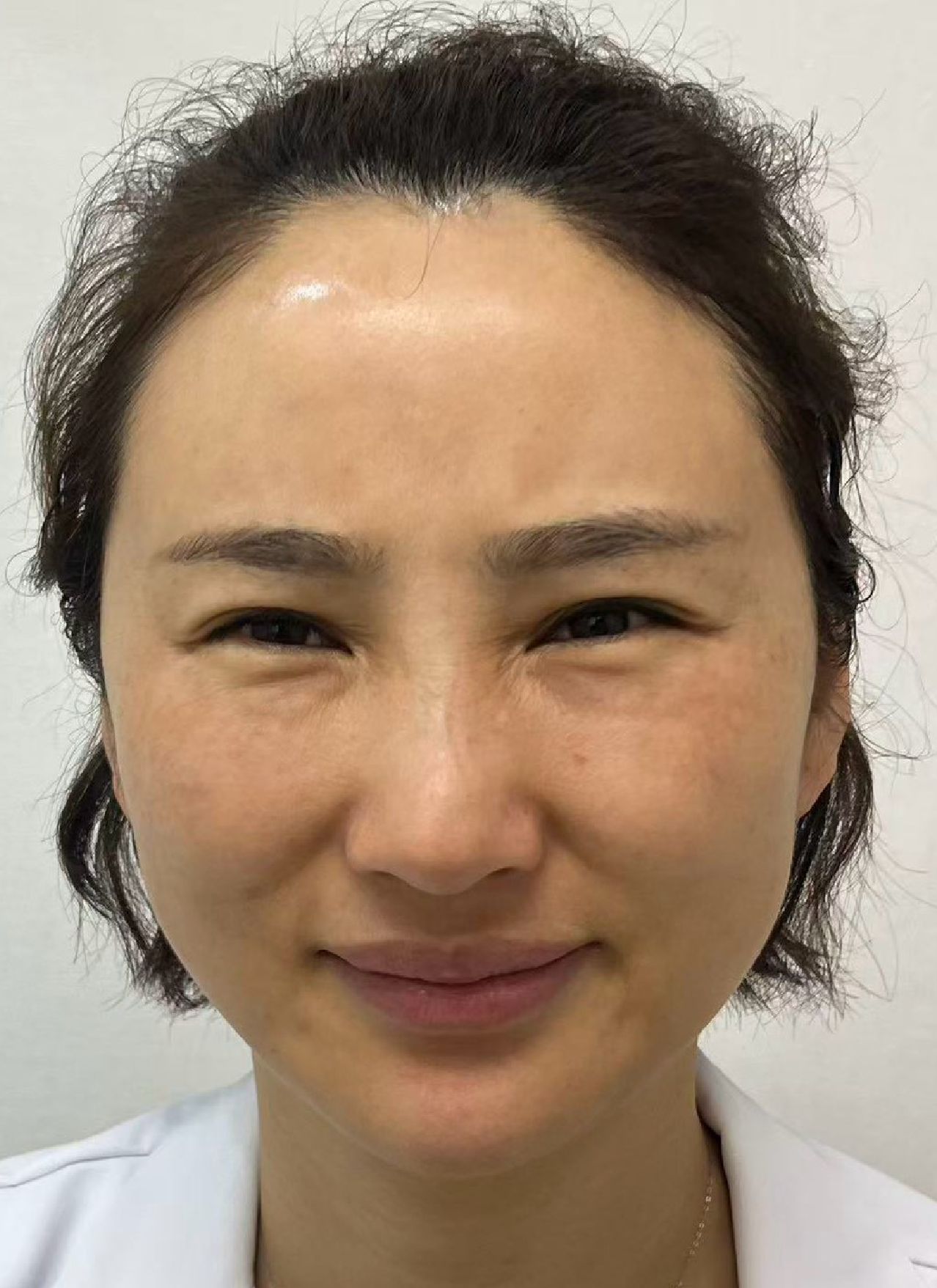
Phase 1 yielded multiple deposits of different materials, including degraded old HA masses, Ellansé nodules, and Sculptra particles. Some materials showed significant fibrotic tissue reaction.
FILLER REVISION Case Notes
FOS cases are among the most complex that the FILLER REVISION team handles, and this case exemplifies why a staged, systematic approach is essential. With three different filler types coexisting across nearly every injection zone, a single-session "clear everything" approach would have been unsafe and counterproductive. The FILLER REVISION protocol for severe FOS involves creating a detailed ultrasound filler map during the first consultation, then prioritizing treatment zones based on which areas contribute most to the unnatural appearance. Each phase is followed by a recovery period and reassessment. This patient-centered pacing is critical — it manages surgical stress, controls swelling, and allows both the physician and the patient to evaluate progress before deciding the next step.
Key Patient Notes
Special Characteristics of FOS Revision
← Swipe to see more →
| Characteristic | Explanation |
|---|---|
| Not a one-time fix | FOS revision almost always requires staged treatment |
| Requires patience | Sessions are usually spaced 3 to 6 months or more apart |
| Progressive improvement | Each phase shows improvement, but not instant transformation |
| Face will undergo transition | Swelling resolution and tissue remodeling take time |
| Psychological preparation matters | Be ready for a lengthy recovery journey |
Recovery Expectations
← Swipe to see more →
| Timeline | Expected Condition |
|---|---|
| Early days after each session | Swelling and bruising are most noticeable |
| About 2–3 weeks | Swelling and bruising mostly settled; the pinhole heals in about 2 weeks |
| About 3 months | That session's final result, varying from person to person |
| Whole staged course | With staged treatment, sessions are usually spaced 3 to 6 months or more apart |
After extraction there is a period in which the swelling settles and firmness gradually softens; we go through the details at your consultation.
Key Insight: FOS revision is a "marathon," not a "sprint." Patients must understand this and maintain realistic expectations for each phase. Impatience may lead to unnecessary risks.
Clinical Takeaways
- Prevention over cure for FOS — regularly review cumulative injection volume; avoid overfilling
- Full-face ultrasound is foundational for FOS management — a complete filler map is essential
- Staged strategy is safe and effective — do not attempt to resolve everything in one session
- Mixed materials increase complexity — different strategies needed for different materials
- Psychological support is essential — the revision journey is long; patients need thorough communication and support
If years of filler injections have left your face looking puffy, distorted, or simply not like you anymore, the FILLER REVISION team understands what you are going through. Our staged FOS revision protocol aims to improve things as much as can be done safely and move you back toward natural facial contours — one step at a time, guided by ultrasound at every stage. We can't guarantee 100% removal.
Related reading:
Frequently Asked Questions
Can pillow face (FOS) be fixed in a single procedure?
In most cases, no. FOS revision almost always requires staged treatment across multiple sessions, because attempting to remove everything at once is unsafe and produces excessive swelling. The approach is to prioritize the most prominent areas first, then address secondary and deeper deposits in later phases, with reassessment in between. We can't guarantee 100% removal; the goal is as much improvement as can be achieved safely, and a return to natural contours.
I already dissolved some of my filler — why is my face still puffy?
Dissolving only works on hyaluronic acid (HA). Materials like Ellansé and Sculptra are non-dissolvable, so they remain in the tissue even after HA dissolution and continue contributing to the puffy appearance. These require physical micro-extraction rather than dissolving, which is why partial HA dissolution often shows only limited improvement.
How long does the whole FOS revision journey take?
With staged treatment, sessions are usually spaced 3 to 6 months or more apart, so the whole course takes longer than a single session. This is because FOS revision is staged, and swelling resolution and tissue remodeling take time. Each phase shows progressive improvement rather than an instant transformation, so psychological preparation for a gradual process is important.
What is the first step before any filler is removed?
The essential first step is full-face ultrasound mapping, which creates a complete inventory of the filler — identifying material types, their locations, and any tissue reactions before any intervention begins. This matters especially when several materials are layered at different depths, because HA areas can be treated differently from non-dissolvable materials. The result is a detailed filler map that guides which zones to prioritize.
What does recovery look like after the first procedure?
The pinhole is small, but this is an extraction, so swelling and bruising are expected, and patience is needed. Swelling and bruising usually settle in about 2–3 weeks, the pinhole heals in about 2 weeks, and that session's final result takes about 3 months, varying from person to person. With staged treatment, sessions are usually spaced 3 to 6 months or more apart.