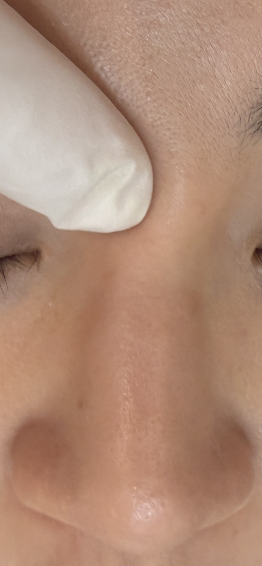
That Puffiness Under Your Eyes—Is It Aging or a Filler Problem?
"I had tear trough filler a year ago, and now I have puffiness under my eyes that looks like eye bags. I tried dissolving it but the puffiness didn't change — did the filler migrate, or is this just aging?" At FILLER REVISION, under-eye puffiness after tear trough filler is one of our most common diagnostic puzzles. Patients arrive after dissolution attempts that may have dissolved native tissue rather than filler, or after being recommended eye bag surgery when the actual cause was migrated filler. In our experience, the answer directly determines what you should do — and getting it wrong means wrong treatment.
True Eye Bags vs. Filler-Induced Pseudo Eye Bags
← Swipe to see more →
| Feature | True Eye Bags (Aging) | Pseudo Eye Bags (Filler Migration) |
|---|---|---|
| Cause | Orbital septum laxity, orbital fat prolapse | Filler migration from tear trough or mid-cheek to the eye bag zone |
| Progression | Gradual over years | May appear months after injection |
| Texture | Soft, more obvious looking upward | Firmer, palpable filler mass |
| Symmetry | Usually roughly symmetric | May be notably asymmetric |
| Dynamic variation | Worsens with fatigue, fluid retention | Relatively fixed, independent of body state |
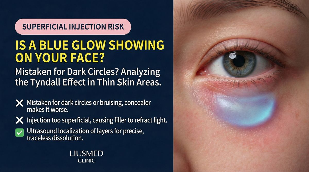
| Skin color | May show darkening | May show Tyndall effect blue translucency |
| Treatment | Eye bag surgery (fat repositioning/removal) | Filler removal |
Key Insight: At FILLER REVISION, we see this pattern regularly — if you surgically treat filler-induced pseudo eye bags as if they were true eye bags, not only does it fail to solve the problem (the bulge is filler, not fat), but the surgeon may encounter unexpected filler material. Conversely, if you inject hyaluronidase for true eye bags thinking it is a filler problem, you dissolve native tissue rather than filler.
Ultrasound (Ultrasonography)—Answers in 10 Minutes
In the under-eye region, ultrasound clearly differentiates orbital fat (specific echo pattern distinct from filler), filler material (each type has unique ultrasound characteristics), tissue edema (clearly distinguishable from filler aggregates), and tear trough ligament position and tension.
This is why ultrasound assessment is our first step for under-eye issues—because a wrong diagnosis leads to wrong treatment.
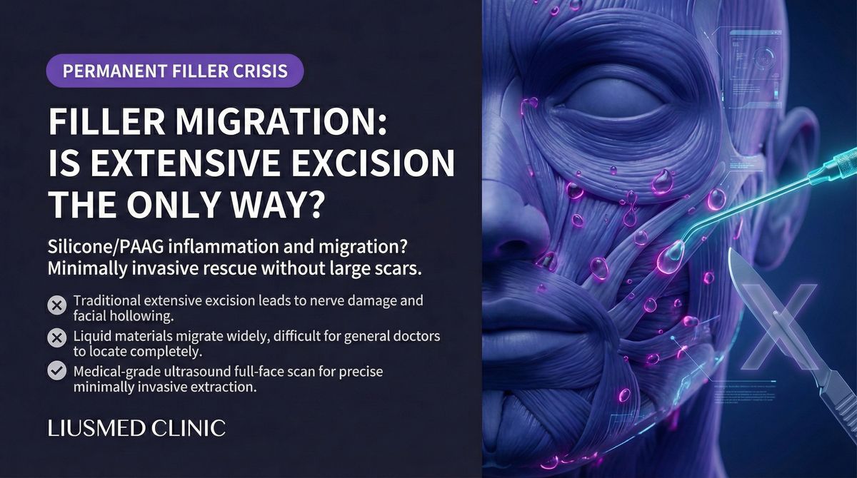
When Filler Migration (Filler Migration) Is Confirmed
Treatment depends on filler type and condition:
- HA (Hyaluronic Acid) (no capsule): Ultrasound-guided precise hyaluronidase
- HA (encapsulated): Ultrasound-guided pinhole extraction
- Non-HA filler: Ultrasound-guided pinhole extraction
- Combined with true eye bags: Address filler first, then assess further needs
The periorbital region is one of the most delicate areas of the face. Our ultrasound-guided pinhole extraction is particularly valuable here—ultrasound reveals the precise structures (nerves, vessels, muscles) in real time, ensuring no critical structures are damaged during extraction.
When Misdiagnosis Leads to Wrong Treatment: The FILLER REVISION Approach
The under-eye area is uniquely prone to diagnostic error because filler-induced pseudo eye bags and true aging eye bags look remarkably similar on visual examination. Patients who reach FILLER REVISION have often undergone the wrong treatment based on this misidentification: dissolution that removed native tissue when the puffiness was actually orbital fat prolapse, or eye bag surgery recommended when the bulge was actually migrated filler. At FILLER REVISION, ultrasound eliminates this diagnostic uncertainty in minutes. The imaging clearly distinguishes orbital fat (with its characteristic echo pattern) from filler deposits (each type producing a unique ultrasound signature), revealing not just what is causing the puffiness but exactly where the material sits. This precision diagnosis then guides the correct treatment — ultrasound-guided extraction for migrated filler, or appropriate referral for true orbital fat issues — ensuring patients receive the right intervention the first time.
Preventing Tear Trough Filler Complications
- Volume is key: The tear trough zone is extremely thin; 0.1ml difference can cause problems
- Choose the right product: Soft, low-hydrophilic products suit the periorbital area
- Correct injection plane: Supraperiosteal is the safest plane
- Physician expertise: Periorbital injection requires deep knowledge of orbital anatomy
- Manage expectations: Tear troughs are not solved by "filling up"; over-filling creates under-eye puffiness
Key Insight: Tear trough filling appears simple but is one of the most technically demanding filler procedures. Thin skin, small space, dense neurovascular structures, and highly visible filler placement errors—these characteristics amplify any injection deviation. See also Tyndall effect in tear troughs and skin atrophy after injection.
Not Sure? Come for a Scan
If you've already tried treatment for under-eye puffiness without success — dissolution that didn't change the puffiness, or a recommendation for surgery you're uncertain about — FILLER REVISION specializes in exactly these cases. Our ultrasound provides the definitive answer in 10 minutes.
Frequently Asked Questions
Is the puffiness under my eye filler or just eye bags?
They look similar but need different treatments, which is why ultrasound matters. Migrated or water-retaining tear-trough filler sits as a defined layer that ultrasound can map; true eye bags are herniated fat with no filler signal. Treating one as the other — for example, doing eye-bag surgery when the cause is filler — leaves the problem unsolved.
Can tear-trough filler cause puffiness years after it was injected?
Yes. Hyaluronic acid in the tear trough can hold water and slowly spread or shift over months to years, producing a soft, sometimes bluish fullness long after the original injection. Because it persists far longer than most people expect, "old" filler is a common hidden cause of new under-eye puffiness.
I had eye-bag surgery but the puffiness is still there — what now?
If puffiness remains or returns after eye-bag surgery, residual or migrated filler is a frequent reason. Ultrasound can show whether filler is still present and where, so the next step targets the actual cause instead of repeating fat-focused surgery.