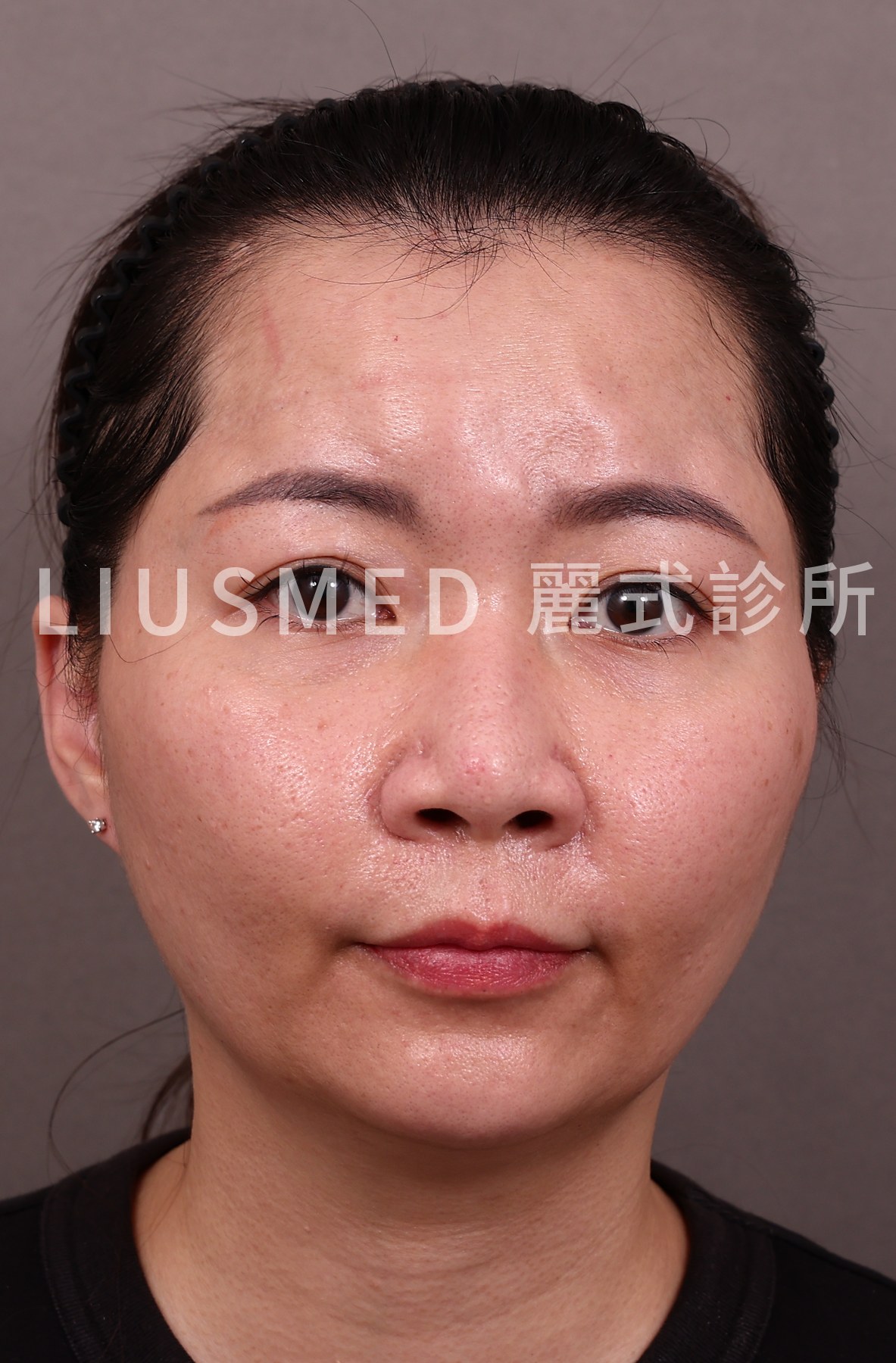
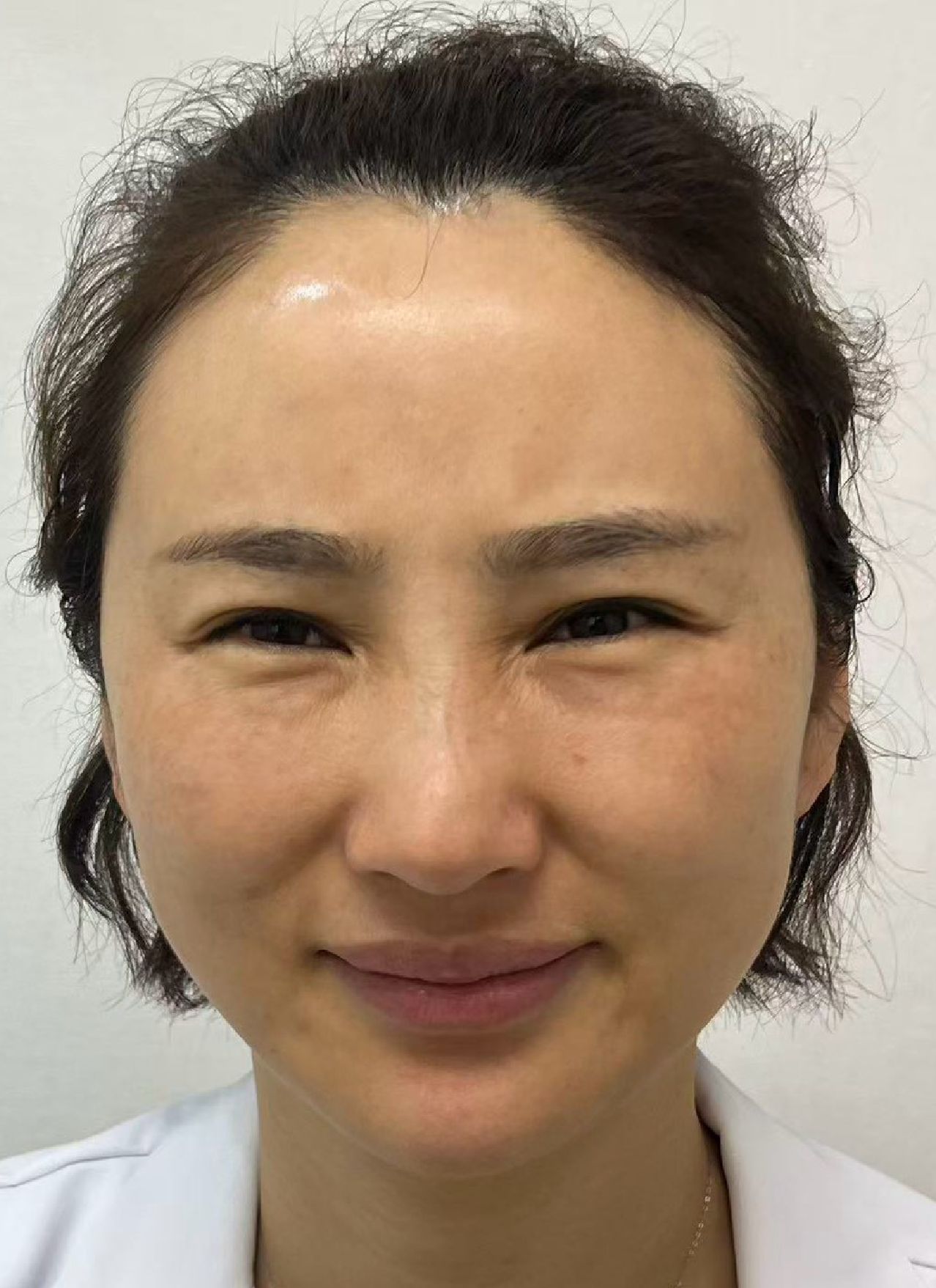
"I Just Had a Radiofrequency Treatment—Why Is My Face Distorted?"
"I had Thermage last month and now the filler in my cheeks looks distorted — nobody warned me this could happen." At FILLER REVISION, we are seeing a growing number of patients whose filler problems were triggered not by the original injection, but by a subsequent energy-device treatment. These cases are particularly frustrating because the patient did nothing wrong — they simply were not told that laser or RF (Radiofrequency) treatments could interact with existing filler.
The root cause is that energy-based devices (lasers, radiofrequency, ultrasound lifting) generate thermal energy that can directly affect fillers within the body. At FILLER REVISION, we assess these energy-device complications with the same ultrasound-first protocol we use for all filler problems — because the damage needs to be seen before it can be repaired.
How Energy Devices Affect Fillers
The Basics of Thermal Effects
Lasers, radiofrequency (RF), and ultrasound lifting devices (such as Ultherapy) share a common mechanism: delivering energy to skin and subcutaneous tissue to produce thermal effects that stimulate collagen remodeling.
- Radiofrequency (RF): Heats the dermal layer to 60-70°C
- Ultrasound (Ultrasonography) lifting: Focused heating to 60-70°C at deeper layers
- Lasers: Heating range from epidermis to dermis depending on wavelength
These temperatures produce controlled effects on normal tissue, but fillers have different physical properties than human tissue and respond differently to thermal energy.
How Different Fillers Respond to Heat
← Swipe to see more →
| Filler Type | Heat Sensitivity | Possible Thermal Damage | Risk Level |
|---|---|---|---|
| Hyaluronic acid (HA) | High | Accelerated degradation, liquefaction, displacement | Moderate-high |
| Calcium hydroxylapatite (Radiesse) | Moderate | Carrier gel liquefaction, microsphere aggregation | Moderate |
| Ellanse (PCL) | Moderate-high | Carrier degradation, altered microsphere behavior | Moderate-high |
| Silicone | Low | Chemically stable but surrounding tissue response changes | Moderate |
| PMMA (Polymethyl Methacrylate) | Low | Microspheres stable but carrier and surrounding tissue affected | Moderate |
| Fat grafting | High | Partial necrosis, liquefaction | High |
Key Insight: At FILLER REVISION, we have observed that even when a filler's chemical structure is unaffected by heat, thermal reactions in surrounding tissue cause indirect problems. Heat-induced local vasodilation, tissue edema, and inflammatory responses can all alter filler position and condition — which is why we always check for filler presence before recommending any energy-based treatment.
Specific Clinical Risks
Risk 1: Filler Softening and Displacement
Thermal energy accelerates cross-link breakdown in certain fillers (especially HA), causing softening. Softened filler is more susceptible to displacement under changes in surrounding tissue pressure.
For more on filler displacement mechanisms: Why Fillers Migrate.
Risk 2: Uneven Degradation Causing Appearance Irregularities
Thermal energy distribution within tissue is not perfectly uniform. Filler may be partially degraded in some areas while remaining intact in others, turning previously smooth augmentation into an uneven surface.
Risk 3: Triggering Encapsulation Reactions
Applying thermal stimulation to areas containing existing fillers may activate local immune responses, promoting fibrosis and capsule formation. This can create new problems around previously stable filler.
For detailed analysis of encapsulation: Encapsulation: Why Dissolvers Fail.
Risk 4: Increased Infection Risk
If low-grade biofilm infection already exists around filler (the patient may be unaware), thermal stimulation may disrupt the existing equilibrium, causing latent infection to acutely flare.
Risk 5: Deep Tissue Injury
Focused ultrasound devices can reach the SMAS (Superficial Musculoaponeurotic System) layer (4.5mm). If filler exists at that depth, focused high temperatures can cause severe localized tissue damage.
← Swipe to see more →
| Energy Device | Primary Risk | Depth of Effect | Fillers of Greatest Concern |
|---|---|---|---|
| RF (Thermage, etc.) | Softening, displacement | Dermis to superficial subcutaneous | HA (Hyaluronic Acid), fat grafting |
| Ultrasound lifting (Ultherapy, etc.) | Deep tissue injury | Dermis to SMAS layer | All deep-layer fillers |
| Fractional laser | Surface degradation | Epidermis to superficial dermis | Superficial HA |
| Intense pulsed light (IPL) | Local heating | Epidermis to superficial dermis | Lower risk |
| Picosecond laser | Minimal thermal effect | Epidermis to dermis | Lower risk but not zero |
Why Many People Only Learn About These Risks After Something Goes Wrong
Incomplete Medical History
Many pre-treatment questionnaires for energy devices do not ask about filler injection history, or ask only about "recent injections" while ignoring treatments from years ago.
Patient Memory Gaps
Many patients do not remember what material was injected years ago or have even forgotten they received injections at all. This is particularly common after "medical tourism."
Practitioner Knowledge Gaps
Some practitioners are unaware of the potential effects of energy devices on fillers, assuming filler injections and light/energy treatments do not interact.
Key Insight: "Not knowing there is filler in the body" does not mean "there is no filler in the body." Many fillers—especially Radiesse, Ellanse, and silicone—persist far longer than patients expect. Material injected years or even a decade ago may still be in place.
The FILLER REVISION Approach: Assessing and Repairing Energy-Device Damage
When patients arrive at FILLER REVISION with filler complications triggered by laser or RF treatments, our first step is never to guess what happened — it is to look. High-resolution ultrasound reveals whether the filler has softened, displaced, fragmented, or triggered an encapsulation response. This imaging also identifies whether underlying issues like biofilm were activated by the thermal stimulus. Based on these findings, we develop a targeted repair strategy: removing displaced or damaged filler through ultrasound-guided extraction while preserving stable, unaffected material. For patients who were never warned about the interaction between energy devices and fillers, this diagnostic clarity is the first step toward restoring both their appearance and their confidence in the treatment process.
What Should Be Done Before Treatment
Comprehensive Treatment History Review
Before any energy device treatment, detailed inquiry should cover:
- All previously injected sites and timing
- Material injected (if known)
- Dosage and depth of injection (if known)
- Whether any existing lumps or abnormalities are present
The Necessity of Ultrasound Examination
For patients with injection history but uncertain about residual status, ultrasound can:
- Confirm whether filler material remains
- Identify filler type and location
- Assess whether encapsulation or other abnormalities exist
- Provide a safety reference for subsequent energy treatments
For more on the ultrasound evaluation process: Filler Repair Evaluation Process.
What to Do If Energy Treatment Has Already Been Performed Over Fillers
If energy device treatment has already been performed on an area containing filler and problems have emerged:
- Stop further treatments—do not continue using energy devices in that area
- Document symptom changes—photograph appearance changes, swelling, texture alterations
- Seek professional evaluation—ultrasound examination to confirm filler status
- Determine management based on findings—may include observation, medication, or filler removal
Schedule a consultation for professional evaluation and the most appropriate management plan.
Frequently Asked Questions
Can I have laser or RF treatment if I already have filler in my face?
It depends on the filler type, depth, and the device — which is why an assessment first matters. Heat-based devices (RF, some lasers, thermage-type treatments) can warm the tissue enough to change or displace certain fillers, while others are relatively unaffected. Knowing what is in your face and where lets the energy treatment be planned safely rather than guessed.
Does laser or RF heat actually melt or move filler?
Heat does not "melt" filler the way many fear, but it can soften, redistribute, or trigger inflammation around some materials — hyaluronic acid behaves differently from collagen stimulators or permanent fillers. The result can be contour change, lumps, or swelling, which is what we assess with ultrasound before deciding on repair.
My filler changed after a laser or RF session — can it be fixed?
Usually yes. The first step is mapping with ultrasound to see whether the material has shifted, clumped, or become inflamed. From there the plan may involve dissolving, precise extraction, or settling the inflammation — chosen according to the filler type and what the energy device actually did.
Conclusion
If your filler has been damaged by a laser, RF, or ultrasound lifting treatment, FILLER REVISION specializes in assessing and repairing these energy-device complications. Our ultrasound evaluation reveals exactly what happened to the filler — displacement, fragmentation, encapsulation, or inflammatory activation — and guides a precise repair plan tailored to your specific situation.
If you have already experienced filler distortion after an energy-device treatment, do not delay assessment. Book a consultation →