Forehead and Temples: High-Risk Revision Zones
"I was told the filler in my temples is too dangerous to remove — that no one would touch it." Patients arrive at FILLER REVISION with this story more often than you might expect. Many clinics decline forehead and temple revision cases outright because of the vascular risks involved. But declining treatment is not the same as the treatment being impossible.
The forehead and temples have seen rapid growth in filler volume in recent years, but they are also among the highest-risk areas for revision. These zones share common characteristics: rich deep vasculature, low surface visibility, and elevated filler migration risk.
Anatomical Specifics
← Swipe to see more →
| Structure | Forehead | Temple |
|---|---|---|
| Skin thickness | Moderate | Thinner |
| Major arteries | Supraorbital, supratrochlear | Superficial temporal artery (STA) and branches |
| Major nerves | Supraorbital, supratrochlear | Temporal branch of facial nerve |
| Common filler planes | Subcutaneous to supraperiosteal | Superficial temporal fascia to deep temporal fascia |
| Migration (Filler Migration) direction | Downward to glabella, lateral to temples | Downward to zygomatic arch |
| Surgical risk level | Medium-High | High |
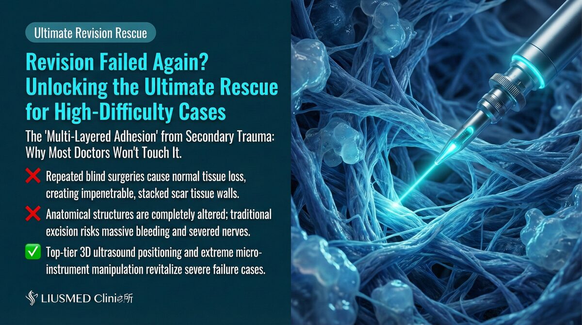
Key Insight: At FILLER REVISION, we've developed specific safety protocols for temple extraction through extensive case experience. The temple is one of the highest-risk zones for filler revision on the entire face. The superficial temporal artery runs superficially with dense branching, and any surgical maneuver must confirm vessel position under ultrasound guidance — a capability central to our approach.
Common Forehead Filler Complications
← Swipe to see more →
| Problem | Presentation | Common Cause |
|---|---|---|
| Migration to glabella | Glabellar elevation or irregularity | Gravity and muscle contraction |
| Surface irregularity | Uneven forehead contour | Inconsistent injection depth |
| Lump formation | Palpable firm nodule | Filler aggregation or fibrosis |
| Lateral migration | Spread from forehead center toward temples | Natural flow of liquid filler |
For more on forehead filler migration, see Forehead Filler Migration and Bumps.
Special Risks of Temple Filler
The Critical Role of the Superficial Temporal Artery
← Swipe to see more →
| Feature | Description |
|---|---|
| Location | On the surface of or within the superficial temporal fascia |
| Course | Curves upward from the preauricular area toward the vertex |
| Branches | Frontal and parietal branches |
| Ultrasound (Ultrasonography) appearance | Pulsatile tubular structure with bright color Doppler signal |
| Surgical significance | Injury can cause major hemorrhage difficult to control with compression |
Temple's Unique Fascial Architecture
The temple has a unique layered structure unlike other facial areas:
- Skin
- Subcutaneous tissue (containing STA, temporal branch of facial nerve)
- Superficial temporal fascia
- Superficial temporal fat pad
- Deep temporal fascia
- Temporalis muscle
Filler may reside in any of these layers, and different materials tend to distribute at different planes.
Ultrasound-Guided Extraction Strategies
Forehead Extraction Key Points
← Swipe to see more →
| Step | Operation | Precaution |
|---|---|---|
| Scanning | Complete scan from forehead to glabella | Confirm any migration |
| Vessel marking | Mark supraorbital artery position | Use color Doppler |
| Incision design | Hidden at hairline or glabella | Select most concealed location |
| Extraction | Layer-by-layer, superficial to deep | Continuous ultrasound monitoring |
| Verification | Ultrasound confirmation of clearance | Watch for deep supraperiosteal residual |
Temple Extraction Key Points
← Swipe to see more →
| Step | Operation | Precaution |
|---|---|---|
| Scanning | Complete scan from temple to zygomatic arch | Track STA (Superficial Temporal Artery) course |
| STA marking | Precisely mark main trunk and branches | Keep color Doppler continuously active |
| Incision design | Hidden within the hairline | Maintain safe distance from STA |
| Extraction | Extremely cautious layered operation | Full-procedure color Doppler monitoring |
| Hemostasis check | Careful post-op confirmation of no bleeding | Temple hematoma has serious consequences |
Key Insight: Temple extraction safety depends entirely on ultrasound real-time guidance. In this zone, operating without ultrasound is effectively blind surgery — an unacceptable risk.
Why FILLER REVISION Confidently Operates Where Others Refuse
The reason most clinics decline forehead and temple cases is simple: without continuous ultrasound guidance, the risk of arterial injury during blind extraction is unacceptably high. At FILLER REVISION, we eliminate this uncertainty by maintaining real-time Color Doppler monitoring throughout the entire procedure. Every instrument movement is tracked relative to the superficial temporal artery and its branches. Our physicians have refined their hairline-concealed incision techniques specifically for these zones, achieving complete extraction through access points that heal virtually invisibly. This combination of vascular mapping, real-time guidance, and concealed-incision design is what allows us to safely treat cases that other clinics consider too risky.
Post-Dissolution Temple Hollowing
Some patients develop severe hollowing after attempting to dissolve temple HA (Hyaluronic Acid). See Temple Hollowing After Dissolving for more.
Conclusion: At FILLER REVISION, High-Risk Does Not Mean Untreatable
Forehead and temple filler extraction demands thorough understanding of deep anatomy combined with ultrasound guidance for safety. At FILLER REVISION, we have built the protocols, the equipment, and the experience to handle these high-risk zones with confidence. If you have been turned away by other clinics or told your forehead or temple filler cannot be safely removed, we invite you to learn what is actually possible.
Related reading: Filler Lump Extraction Technique
Frequently Asked Questions
Other clinics told me my forehead or temple filler is too dangerous to remove. Does that mean it really can't be done?
Many clinics decline forehead and temple revision cases because operating without ultrasound guidance poses unacceptable vascular injury risks. But declining treatment is not the same as the treatment being impossible. At FILLER REVISION these zones are handled under continuous Color Doppler ultrasound, which lets the vessels be tracked in real time. If you have been turned away, you are welcome to come for a professional ultrasound assessment to learn what is actually possible.
Will the extraction leave a visible scar on my forehead or temple?
The incisions for forehead and temple extraction are designed to be concealed within the hairline, and they heal virtually invisibly. The access point is chosen as the most concealed location while keeping a safe distance from the superficial temporal artery. This concealed-incision design is part of how these high-risk zones are approached safely.
How does the doctor actually find my filler before removing it in the temple?
Filler in the temple region can reside in any of six distinct tissue layers, and different materials tend to settle at different planes. Ultrasound is used first to scan the area, determine the exact plane the filler sits in, and map the course of the superficial temporal artery. Only after this mapping does layer-by-layer extraction begin, with ultrasound confirming how completely the filler has been cleared.
What should I watch for and how should I care for the area after the extraction?
For the forehead, avoid broad pressure on the area and keep your head slightly elevated when sleeping. For the temple, avoid side-sleeping that presses on the area and watch for persistent swelling, which can indicate a hematoma. A temple hematoma can have serious consequences, so careful post-operative checking for any bleeding is part of the procedure, and you should follow up if swelling does not settle.
I developed hollowing in my temple after trying to dissolve the filler. Can that situation be assessed too?
Some patients develop severe hollowing after attempting to dissolve temple HA (hyaluronic acid). The article addresses this scenario and links to dedicated information on post-dissolution temple hollowing. If you are in this situation, you are welcome to be assessed; understanding what happened to the tissue is the starting point for deciding the next step.