Case Scenario
"The clinic told me it was just bruising and to go home. But the skin kept getting darker and the pain wouldn't stop." By the time the referring clinic called FILLER REVISION for an emergency transfer, five hours had passed — and every minute mattered.
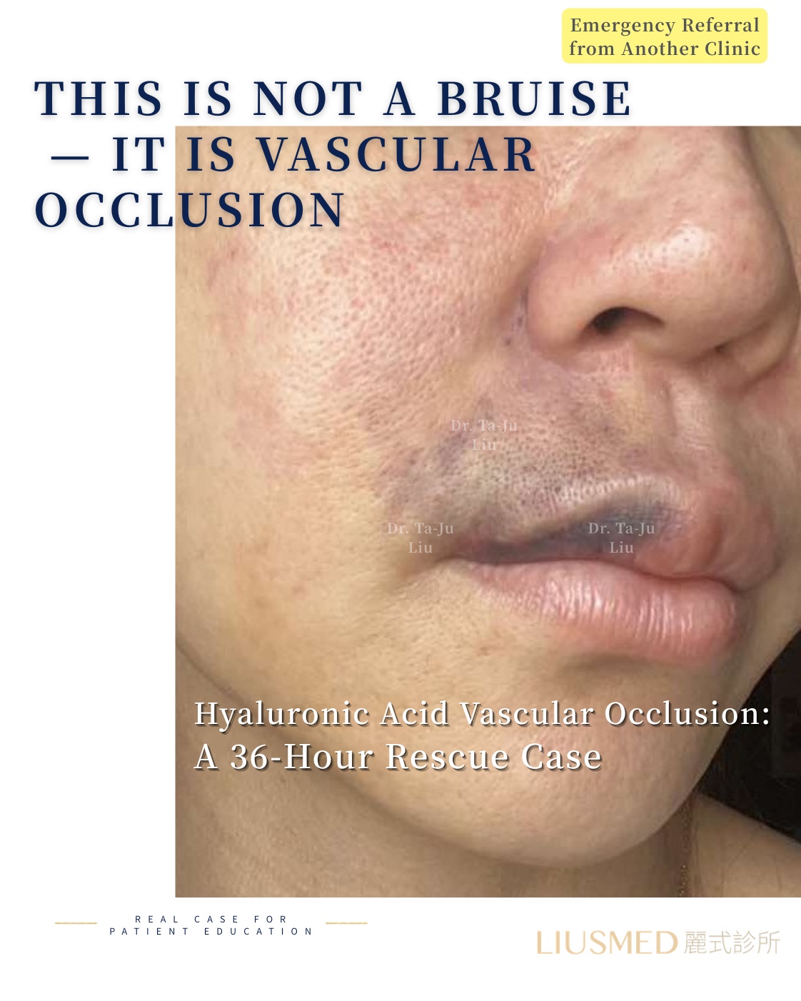
Patient Background: A woman in her 30s who received hyaluronic acid injections in the nasal and nasolabial fold areas at another clinic. During the injection, the patient began experiencing unusual stinging and burning sensations near the injection site. Approximately 20 minutes after the injection was completed, blanching appeared in the perialar region, which progressed to a violaceous discoloration within one hour.
Emergency Transfer Timeline:
- 20 minutes post-injection: Blanching appeared in the perialar region; patient reported escalating local pain
- 1 hour post-injection: Blanched area began turning purplish-red with slight expansion
- Original clinic attempted pressure and warm compress management, but symptoms continued to worsen
- ~3 hours post-injection: Affected area darkened further with increasingly defined borders
- Original clinic contacted our facility for emergency transfer
- ~5 hours post-injection: Upon arrival at our clinic, the affected area had progressed to deep purple to dark discoloration
Presentation on Arrival:
- Sharply demarcated deep purple to dark skin discoloration from the perialar region to the nasolabial fold
- Noticeably reduced skin temperature in the affected zone
- Patient reported persistent pain and numbness in the affected area
- Capillary refill test indicated severely diminished blood flow in the affected region
- Patient in extreme distress and anxiety
Deep Analysis
Root Cause Analysis
← Swipe to see more →
| Aspect | Finding |
|---|---|
| Occlusion mechanism | Filler directly injected into or compressing a branch of the facial artery, interrupting blood flow |
| Affected vessel | Based on ischemic territory distribution, likely involving the alar artery and its branches |
| Time factor | Approximately 5 hours elapsed from symptom onset to rescue initiation — within the critical tissue salvage window |
| Initial ultrasound assessment | Evidence of filler compressing vasculature in the affected zone; significantly diminished blood flow signals |
| Tissue perfusion status | Deep purple to dark discoloration indicating severe ischemia, but not yet progressed to irreversible necrosis |
Key Insight: Vascular occlusion is one of the most serious acute complications of filler injection. Time is the critical determinant of tissue survival — the earlier intervention begins, the greater the probability of complete tissue recovery. Classic warning signs include: abnormal pain during injection, blanching or purplish discoloration around the injection area, and decreased skin temperature. Once these signs appear, emergency protocols should be initiated immediately rather than adopting a wait-and-see approach. FILLER REVISION maintains emergency rescue readiness specifically for these time-critical situations, where ultrasound-guided precision hyaluronidase delivery can mean the difference between full recovery and permanent tissue damage.
Related reading: Vascular Occlusion Mechanism and Emergency Response
Doctor's Perspective
"The timing of this patient's transfer was critical. While 5 hours is not the ideal intervention window — the earlier the better — it was still within the time frame where tissue salvage remained possible. The deep purple to dark skin color indicated severe ischemia, but through ultrasound assessment, we determined that the tissue had not yet progressed to complete necrosis, meaning a window of opportunity for rescue still existed.
Our emergency strategy was multi-pronged: first, immediately perform ultrasound-guided precision localization to confirm the exact position of the filler and its compression effect on surrounding vasculature; second, administer high-concentration hyaluronidase under ultrasound guidance, delivering it directly to the filler material compressing the vessel; third, implement vasodilation and anticoagulation support measures to facilitate perfusion recovery. The entire rescue process demands both speed and precision — every minute matters for tissue survival."
Treatment Plan and Process
Emergency Strategy
← Swipe to see more →
| Phase | Treatment | Timeline |
|---|---|---|
| Phase 1 | Ultrasound (Ultrasonography) emergency assessment and precision localization | Within 15 minutes of arrival |
| Phase 2 | Ultrasound-guided precision hyaluronidase injection | Immediately following assessment |
| Phase 3 | Vasodilation and perfusion support treatment | Concurrent with hyaluronidase |
| Phase 4 | Continuous monitoring and supplemental treatment | 24-48 hours post initial intervention |
Emergency Rescue Process
- Ultrasound emergency assessment: Rapid scanning to confirm filler location, extent of vascular compression, and residual blood flow status
- Precision hyaluronidase injection: Under real-time ultrasound guidance, high-concentration hyaluronidase delivered directly to the filler compressing the vessel
- Sequential supplemental injections: Based on real-time ultrasound monitoring, additional hyaluronidase administered to areas of persistent compression
- Vasodilation support measures: Local and systemic blood flow enhancement therapy implemented concurrently
- Real-time monitoring: Skin color changes and capillary refill assessed every 15-30 minutes
- Follow-up treatment: 24-hour follow-up evaluation to determine need for additional intervention
Rescue Outcome
Approximately 2-3 hours after the initial emergency intervention, the affected skin color began transitioning from dark to a deep red, indicating blood flow restoration. At the 24-hour follow-up, most of the area had progressed to red to light red, with skin temperature gradually returning to normal. Through several days of intensive monitoring and supportive treatment, tissue perfusion progressively normalized.
FILLER REVISION Emergency Protocol Notes
This case underscores why FILLER REVISION treats vascular occlusion rescue as a distinct clinical discipline, not simply "dissolving filler faster." The multi-pronged strategy — ultrasound-guided precision hyaluronidase delivery, vasodilation support, and continuous perfusion monitoring — requires equipment, training, and protocols that go beyond standard clinic readiness. What made this rescue possible within the critical window was the combination of real-time ultrasound localization of the compression source and the ability to deliver high-concentration hyaluronidase directly to the affected vessel. FILLER REVISION accepts emergency referrals from other clinics for exactly these situations, because rapid access to ultrasound-guided intervention can be the deciding factor in tissue survival.
Key Patient Notes
Special Characteristics of Vascular Occlusion Emergency
← Swipe to see more →
| Characteristic | Explanation |
|---|---|
| Time equals tissue | The earlier intervention begins, the greater the chance of complete tissue recovery |
| Recovery is gradual | After blood flow restoration, tissue repair still requires weeks to months |
| Temporary marks may remain | Skin that experienced severe ischemia may undergo a post-inflammatory hyperpigmentation phase; most improve gradually |
| Intensive follow-up is essential | The first few days after rescue require frequent follow-up visits to monitor progress |
| Psychological support matters equally | Patients who experience an occlusion event often suffer significant psychological trauma and need thorough support and communication |
Recovery Timeline
← Swipe to see more →
| Timeline | Expected Condition |
|---|---|
| Hours after rescue | Skin color transitions from dark to deep red, indicating blood flow restoration |
| Days 1-3 post-rescue | Continued improvement; affected area may exhibit swelling and inflammatory response |
| Weeks 1-2 post-rescue | Acute phase resolves; tissue enters repair phase; possible hyperpigmentation |
| Months 1-3 post-rescue | Tissue continues to heal; hyperpigmentation gradually improves |
| Month 6 post-rescue | Most cases achieve good recovery; assessment of whether cosmetic follow-up treatment is needed |
Key Insight: Successful vascular occlusion rescue does not mean immediate return to normal. Tissue that has experienced ischemia requires time to repair, and the recovery process may include temporary manifestations such as hyperpigmentation and texture changes. The important thing is to maintain regular follow-up so the physician can assess recovery progress and provide further restorative treatment at the appropriate time.
Clinical Takeaways
- Timely recognition of warning signs is life-saving — abnormal pain during injection and blanching or purplish discoloration are early occlusion signals requiring immediate action
- The time window determines outcomes — the first 6 hours after occlusion onset represent the golden period for rescue
- Ultrasound's value in emergency rescue is irreplaceable — precise localization of the compression source enables precise decompression
- Multi-pronged rescue strategy is essential — hyaluronidase, vasodilation, and perfusion support must proceed simultaneously
- Injection safety is fundamental — understanding the vascular anatomy of facial danger zones is the first step in occlusion prevention
If you or a patient is experiencing signs of vascular occlusion — skin blanching, darkening, or severe pain after filler injection — time is critical. FILLER REVISION provides emergency ultrasound-guided rescue services. Do not wait for symptoms to resolve on their own.
Related reading:
- Vascular Occlusion Mechanism and Emergency Response
- Facial Injection Danger Zones
- Filler Repair Evaluation Process
Frequently Asked Questions
After a filler injection, how do I know if I'm having a vascular occlusion rather than just normal bruising?
According to this article, the classic early warning signs of vascular occlusion are abnormal pain during the injection, blanching (whitening) or purplish discoloration of the skin near the injection site, and a decrease in skin temperature in that area. In this patient's case, blanching appeared in the perialar region about 20 minutes after injection and progressed to a violaceous discoloration within one hour. The article stresses that once these signs appear, emergency protocols should be started immediately rather than adopting a wait-and-see approach.
It's already been a few hours since my skin started darkening. Is it too late to be rescued?
Not necessarily. The article describes the first 6 hours after onset as the golden rescue window for tissue salvage, and the earlier intervention begins, the greater the chance of full tissue recovery. In this case, the patient arrived about 5 hours after onset — which the doctor notes is not the ideal window, but was still within the time frame where tissue salvage remained possible because ultrasound assessment showed the tissue had not yet progressed to irreversible necrosis. The article advises not to wait for symptoms to resolve on their own and to seek emergency care quickly.
Why does this clinic use ultrasound during the rescue instead of just injecting the dissolving agent?
The article explains that ultrasound-guided precision hyaluronidase delivery directly to the compressing filler is far more effective for emergency decompression than blind injection. The ultrasound first confirms the exact filler location, the extent of vascular compression, and residual blood flow, so the high-concentration hyaluronidase can be delivered straight to the filler that is pressing on the vessel. The article describes this real-time ultrasound localization as having an irreplaceable value in emergency rescue, because precise localization of the compression source enables precise decompression.
If the rescue works, will my skin go back to normal right away?
No. The article is clear that a successful rescue does not mean an immediate return to normal. Even after blood flow is restored, full tissue recovery takes weeks to months, and the skin that experienced severe ischemia may go through a temporary post-inflammatory hyperpigmentation phase that most often improves gradually. In this case, the article describes a recovery timeline where most cases achieve good recovery by around 6 months, at which point the physician assesses whether any cosmetic follow-up treatment is needed.
My filler was injected at a different clinic. Can I still come here for emergency help?
Yes. The article describes this exact situation: the patient received filler at another clinic, and when symptoms worsened the original clinic contacted FILLER REVISION for an emergency transfer. The article states that FILLER REVISION accepts emergency referrals from other clinics for exactly these time-critical situations, because rapid access to ultrasound-guided intervention can be the deciding factor in tissue survival. If you have skin blanching, darkening, or severe pain after a filler injection, the article advises that time is critical and not to wait for symptoms to resolve on their own.
I'm very shaken after going through this. Will I get any support beyond the physical treatment?
Yes. The article explicitly notes that psychological support matters equally, because patients who experience an occlusion event often suffer significant psychological trauma and need thorough support and communication. Alongside the physical rescue and recovery monitoring, the article frames this support and communication as part of the care. It also emphasizes intensive follow-up in the first few days after rescue and regular follow-up visits so the physician can assess recovery progress over time.