Why Is "Revising a Revision" So Difficult?
"My first revision made things worse. Now I have more scarring, more fibrosis, and no one wants to touch it." At FILLER REVISION, secondary revision cases are not exceptions — they are a significant portion of our practice. When a filler revision surgery fails to achieve the expected outcome — or even creates new problems — the patient needs a secondary revision. This is widely recognized as the most challenging work in the filler revision field.
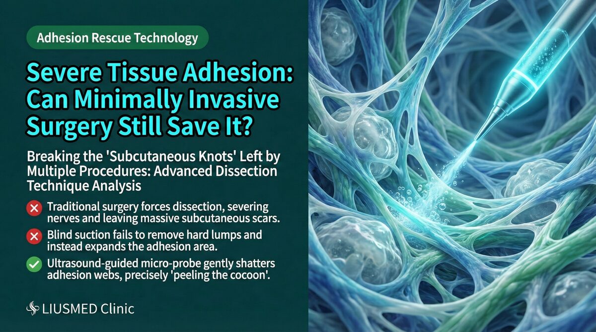
The reason is straightforward: the first revision has already altered the original tissue architecture. Fibrosis is more severe, anatomical landmarks may have been destroyed, and scar tissue has obscured once-clear tissue planes — all of this causes secondary revision difficulty to increase exponentially.
Key Insight: At FILLER REVISION, we've developed specialized protocols for exactly these scenarios. Secondary revision difficulty is not twice that of primary revision — it may be several times greater. Every surgery leaves traces in the tissue, and these traces progressively narrow the operating space and raise the risk profile for each subsequent procedure. This is why secondary revision demands the highest level of ultrasound expertise.
Unique Challenges of Secondary Revision
Tissue-Level Changes
← Swipe to see more →
| Change | Impact | Increased Risk |
|---|---|---|
| Worsened fibrosis | Tissue becomes hard, loses elasticity | Extraction difficulty increases |
| Scar formation | Surgical pathways blocked by scar | New operative routes must be found |
| Vascular changes | Original vessels may be damaged or displaced | Bleeding risk increases |
| Neural changes | Nerve pathways may be altered | Nerve injury risk increases |
| Plane obliteration | Previously clear anatomical planes destroyed | Precise operation becomes more difficult |
| Tissue deficiency | Depressions from over-extraction | Reconstruction becomes more complex |
Filler-Level Changes
← Swipe to see more →
| Change | Description |
|---|---|
| Residual filler | Portions not fully removed in the first surgery |
| Fragmentation | Filler becomes scattered after partial removal |
| Deep displacement | Surgical manipulation may push filler to deeper layers |
| Mixed materials | Different materials from different injection sessions may coexist |
| Thicker encapsulation | Residual filler enclosed by denser fibrous tissue |
Common Types of Revision Failure
Why Does the First Revision Fail?
← Swipe to see more →
| Failure Type | Cause | Outcome |
|---|---|---|
| Incomplete clearance | No ultrasound guidance; deep residuals missed | Problem persists or recurs |
| Over-extraction | Excessive removal causing tissue deficiency | Depression, asymmetry |
| New iatrogenic damage | Vascular or nerve injury during operation | New complications |
| Wrong plane | Operating in the incorrect tissue layer | Normal tissue destroyed |
| Improper dissolution | Non-selective dissolution affecting normal tissue | Depression, unevenness |
| Infection | Surgical site infection | Further tissue damage |
Key Insight: Most revision failures trace back to two root causes: operating without ultrasound guidance or insufficient revision experience. Operating without visualization is effectively blind surgery.
Ultrasound (Ultrasonography) Assessment for Secondary Revision
Why Secondary Revision Needs Ultrasound Even More
In tissue already altered by surgery, the importance of ultrasound is amplified to its maximum:
← Swipe to see more →
| Assessment Need | Information Ultrasound Provides |
|---|---|
| Residual filler | Precise location of filler missed in the first surgery |
| Fibrosis extent | Evaluation of fibrosis range and severity |
| Scar distribution | Confirmation of scar tissue location and extent |
| Current vascular status | Verification of post-surgical vessel courses |
| Tissue deficiency | Assessment of depression from over-extraction |
| Normal tissue | Identification of remaining normal tissue structures |
Pre-Operative Ultrasound Assessment Workflow
← Swipe to see more →
| Step | Content | Purpose |
|---|---|---|
| Complete scan | Full scan of surgical area and surroundings | Establish comprehensive current status map |
| Residual localization | Mark positions of residual filler | Plan extraction targets |
| Fibrosis assessment | Evaluate depth and extent of fibrosis | Estimate extraction difficulty |
| Vascular remapping | Re-confirm vessel courses | Update safety roadmap |
| Comparative assessment | Compare with contralateral side or pre-op images | Set revision goals |
| Feasibility judgment | Comprehensive assessment of surgical viability | Determine suitability for re-operation |
Surgical Strategy for Secondary Revision
Strategic Differences from Primary Revision
← Swipe to see more →
| Strategy Item | Primary Revision | Secondary Revision |
|---|---|---|
| Incision choice | Optimal location available | May be limited by prior scars |
| Operating space | Relatively ample | Compressed by fibrosis |
| Extraction difficulty | Standard | Significantly increased |
| Bleeding risk | Standard | Increased (altered vessel courses) |
| Conservative approach | Standard conservative | Even more conservative |
| Staged strategy | As needed | Strongly recommended |
| Ultrasound dependency | High | Extremely high |
Key Surgical Execution Points
- Maximum conservative principle: Better to leave a small residual than risk damaging normal tissue
- Multi-session staged strategy: Nearly all secondary revisions should be divided into 2–3 sessions
- Full-procedure ultrasound guidance: Every operative step performed under ultrasound monitoring
- Real-time strategy adjustment: Immediate strategy modification based on intraoperative findings
- Sufficient recovery intervals: Allow adequate time between sessions for tissue recovery
Key Insight: The golden rule of secondary revision is "small amounts, multiple sessions." Aggressive operation in already-damaged tissue only creates more damage. Staged extraction gives tissue time to recover and allows the physician to reassess between each session.
Why FILLER REVISION Succeeds at Secondary Revision Where the First Attempt Failed
The primary reason first revisions fail is operating without ultrasound — the surgeon cannot see residual filler, altered vessel courses, or scar tissue boundaries. At FILLER REVISION, our approach to secondary revision begins with what the first surgeon lacked: a complete ultrasound reassessment that maps the current tissue state, including all changes caused by the previous surgery. We then apply our "small amounts, multiple sessions" protocol — never attempting aggressive single-session correction in already-damaged tissue. Each session addresses a targeted portion under full ultrasound guidance, with adequate recovery time between sessions for tissue to stabilize. This patient, methodical approach transforms cases that seem hopeless after a failed first attempt into achievable — if gradual — improvements.
Managing Severe Fibrosis
One of the most common challenges in secondary revision is severe fibrosis. For more on fibrosis management, see Severe Adhesion and Fibrosis Extraction.
← Swipe to see more →
| Fibrosis Severity | Management Strategy | Expected Outcome |
|---|---|---|
| Mild | Meticulous separation then extraction | Good |
| Moderate | Requires longer time and more sessions | Acceptable |
| Severe | Partial extraction + long-term follow-up | May need to accept some residual |
| Extreme | Conservative observation primarily | Symptom improvement as goal |
Patient Expectation Management
Realistic Expectations for Secondary Revision
← Swipe to see more →
| Aspect | Realistic Expectation | Unrealistic Expectation |
|---|---|---|
| Complete clearance | Significant improvement with possible small residual | 100% removal of all filler |
| Appearance recovery | Noticeable improvement with possible minor imperfections | Return to perfect pre-injection state |
| Recovery time | Longer than primary revision | Same as primary revision |
| Number of sessions | May require 2–3 sessions | Resolving everything in one session |
| Final results | Assessed at 3–6 months | Immediately visible final outcome |
How to Avoid Needing Secondary Revision
Keys to Successful Primary Revision
- Choose a physician with ultrasound capability: Ultrasound guidance dramatically reduces revision failure rates
- Complete pre-operative assessment: Thorough understanding of the problem enables correct surgical planning
- Experienced revision specialist: The learning curve for revision surgery is steep
- Realistic expectation setting: Thorough communication with your physician about anticipated outcomes
- Proper post-operative care: Following physician instructions for aftercare
Post-Operative Care and Follow-Up
← Swipe to see more →
| Timeline | Special Considerations |
|---|---|
| Weeks 1–2 | Stricter care period, longer than primary revision |
| Month 1 | Evaluate initial recovery; decide if next session needed |
| Month 3 | Interim assessment; tissue beginning to stabilize |
| Month 6 | Evaluate final results; develop follow-up plan |
| Year 1 | Long-term follow-up to confirm stability |
Conclusion: FILLER REVISION Specializes in the Cases Others Cannot Solve
Secondary revision is the most challenging surgery in the filler revision field. Altered tissue architecture, more severe fibrosis, lost anatomical landmarks — all of this demands the highest caliber of ultrasound interpretation ability and surgical skill. At FILLER REVISION, these complex cases are not rare exceptions but a core part of our practice.
If you have experienced a failed revision or are living with complications from a previous repair attempt, do not assume your situation is beyond help. FILLER REVISION has the expertise and equipment to assess even the most complex cases.
Related reading: Severe Adhesion and Fibrosis Extraction, Filler Lump Extraction Technique, Filler Repair Evaluation Process
Frequently Asked Questions
My first filler revision made things worse. Is a second revision even possible, or is my situation beyond help?
A failed first revision does not mean your situation is beyond help. These secondary revision cases are a significant part of FILLER REVISION's practice, not rare exceptions. The approach begins with a complete ultrasound reassessment that maps the current tissue state — including all the changes the previous surgery caused — before any operation. With this groundwork, cases that seem hopeless after a failed first attempt can become achievable, if gradual, improvements.
Why is a second revision harder than the first one?
Because the first revision has already altered the original tissue architecture. Fibrosis is more severe, anatomical landmarks may have been destroyed, and scar tissue has obscured once-clear tissue planes — so the difficulty increases exponentially, not just twice. Each surgery also leaves traces that narrow the operating space and raise the risk for the next procedure. This is why secondary revision demands the highest level of ultrasound expertise.
Why did my first revision fail in the first place?
Most revision failures trace back to two root causes: operating without ultrasound guidance, or insufficient revision experience. Operating without visualization is effectively blind surgery — the surgeon cannot see residual filler, altered vessel courses, or scar tissue boundaries. This is why deep residuals get missed, normal tissue can be damaged, and problems persist or recur.
How many sessions will a second revision take, and when will I see the final result?
You should expect 2–3 sessions with recovery intervals between them, because the golden rule for secondary revision is 'small amounts, multiple sessions.' Aggressive single-session correction in already-damaged tissue only creates more damage, so each session addresses a targeted portion and allows the tissue time to recover and the physician to reassess. The final results are best assessed at 3–6 months after surgery, not immediately.
Can a second revision remove 100% of my filler and restore my original face?
A realistic expectation is significant improvement with the possibility of a small residual — not 100% removal of all filler. Likewise, you can expect noticeable improvement in appearance, though minor imperfections may remain, rather than a return to a perfect pre-injection state. This is by design: the conservative principle is that it is better to leave a small residual than to risk damaging normal tissue. In severe fibrosis cases, accepting some residual may be part of the plan.
Why is ultrasound guidance even more important for a second revision than a first one?
Because the prior surgery has altered your vessel courses, nerve pathways, and tissue planes, so the surgeon cannot rely on normal anatomy. Ultrasound lets the physician precisely locate residual filler the first surgery missed, evaluate the extent of fibrosis and scar distribution, and re-confirm the current vessel courses before operating. Performing every operative step under ultrasound monitoring is how the increased bleeding and nerve-injury risks of altered tissue are kept in check.