Choosing the Right "Eyes" to See Your Filler
If you are dealing with a filler complication — lumps, swelling, asymmetry — and your doctor has suggested imaging, you are probably wondering which method will actually show what is going on inside. Should you get an MRI (Magnetic Resonance Imaging), or is ultrasound better? The answer depends on what you need to learn and what you plan to do next.
The two most commonly used non-invasive imaging tools are MRI (magnetic resonance imaging) and high-resolution ultrasound. Studies have demonstrated ultrasound's particular value in improving the safety and outcomes of filler treatments (Schelke et al., 2018), while imaging of soft-tissue foreign bodies including fillers has been reviewed for both modalities. Each excels in different areas, and understanding their differences helps you make a better choice.
Key Insight: At FILLER REVISION, our clinical experience confirms that MRI and ultrasound are not either/or choices but complementary tools with different indications. For most filler-related clinical problems — localization, distribution mapping, real-time treatment guidance — high-resolution ultrasound is typically the first choice because it enables diagnosis and treatment in a single session.
Basic Principles of Each Modality
MRI
MRI uses powerful magnetic fields and radiofrequency pulses to generate signals from hydrogen nuclei in tissue. Different tissues have different hydrogen densities, producing images with exceptional soft tissue contrast.
High-Resolution Ultrasound (Ultrasonography)
Ultrasound uses high-frequency sound waves (15-50 MHz) reflected from tissue interfaces to create real-time structural images. Different density tissues produce different echo patterns.
Comprehensive Comparison
← Swipe to see more →
| Comparison | MRI | High-Resolution Ultrasound |
|---|---|---|
| Spatial resolution (superficial) | Moderate (0.5-1mm) | Very high (0.1-0.3mm) |
| Spatial resolution (deep) | High (uniform) | Decreases with depth |
| Soft tissue contrast | Very high | Moderate |
| Filler identification | Good (HA bright on T2) | Good (characteristic echoes per material) |
| Real-time capability | No (scheduled, 30-60 min) | Yes (immediate, 5-15 min) |
| Treatment guidance | Not applicable | Real-time injection and extraction guidance |
| Cost | High | Moderate |
| Accessibility | Hospital/imaging center required | Office-based |
| Repeatability | Lower (cost/time constraints) | High (easy repeat follow-up) |
| Contraindications | Metal implants, claustrophobia | Virtually none |
| Full-face scanning | Single session | Region-by-region examination |
Filler Appearance on Each Modality
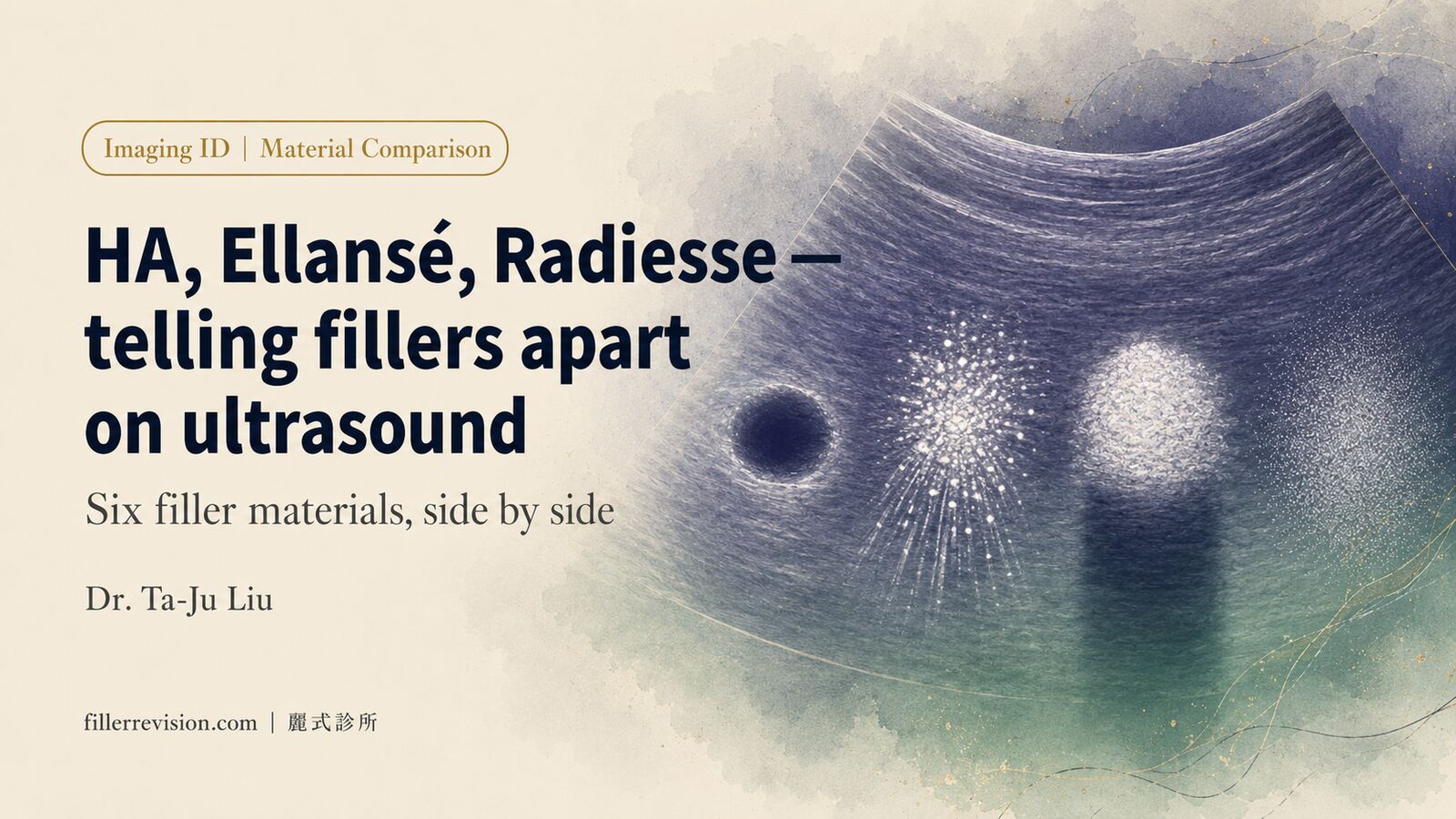
Hyaluronic Acid (HA)
MRI: High water content HA (Hyaluronic Acid) appears bright on T2-weighted images, providing excellent contrast against surrounding tissue. MRI is particularly sensitive for detecting HA residue.
Ultrasound: HA appears as hypoechoic to anechoic round deposits with clear boundaries. Contrast against surrounding edematous tissue may be lower than MRI.
Silicone/Permanent Fillers
MRI: Unique signal characteristics; special silicone-suppression sequences can confirm presence.
Ultrasound: Strong echogenicity with "snowstorm" effect, usually easily identifiable.
Calcium-Based Fillers (CaHA)
MRI: Calcified material appears as low signal, less intuitive than CT (Computed Tomography).
Ultrasound: Strongly hyperechoic with posterior acoustic shadowing, easiest to identify on ultrasound.
How FILLER REVISION Uses Imaging for Revision Planning
For revision patients at FILLER REVISION, the imaging choice has direct consequences for treatment efficiency and outcomes. High-resolution ultrasound serves as our primary assessment tool because it enables a seamless workflow: assess the filler, identify the complication, and — when appropriate — proceed to ultrasound-guided treatment in the same session. This "see and treat" capability eliminates the diagnostic delays that come with scheduling MRI at a separate facility, waiting for interpretation, and then returning for treatment. For patients who have already undergone MRI elsewhere, we integrate those findings with our ultrasound assessment to build a complete picture. The goal is always the same: understand what is inside before deciding how to address it.
Clinical Scenario Selection Guide
When to Choose Ultrasound
- Initial assessment: Screening and localization tool
- Filler repair evaluation: Real-time assessment and treatment planning
- Treatment guidance: Image-guided injection or extraction needed
- Follow-up monitoring: Post-treatment periodic assessment
- Before and after filler extraction: Pre-operative localization and post-operative confirmation
When to Choose MRI
- Deep structure assessment: Suspected deep or widespread filler distribution
- Full-face wide field scanning: Need overall distribution overview when filler location is uncertain
- Specific material confirmation: Need to confirm specific materials like silicone
- Legal or documentation purposes
- When ultrasound assessment is inconclusive
Key Insight: In daily clinical practice, high-resolution ultrasound is the preferred tool for filler assessment due to its real-time capability, high resolution, treatment guidance ability, and accessibility. MRI serves as an important complementary modality in specific situations.
Ultrasound's Unique Advantage: Seamless Connection Between "Seeing" and "Treating"
Ultrasound's most irreplaceable advantage: it is both a diagnostic and treatment guidance tool.
In a single session, the physician can:
- Assess filler location, type, and encapsulation status with ultrasound
- Upon discovering a problem, immediately perform precise treatment under ultrasound guidance
- Confirm treatment results in real time
This instant "diagnose-treat-confirm" workflow is something MRI cannot provide.
Learn more about ultrasound in filler identification. At FILLER REVISION, we use the imaging modality that best serves your specific situation — because the right diagnosis drives the right treatment.
Key Insight: The best imaging tool is not the "most expensive" or "most advanced" — it is the one that best helps solve your problem. For the vast majority of filler-related clinical issues, high-resolution ultrasound delivers optimal value because it enables "seeing" and "treating" to happen at the same time, same place, by the same physician.
Frequently Asked Questions
I have filler lumps and swelling — should I get an MRI or an ultrasound?
For most filler-related problems — localization, distribution mapping, and real-time treatment guidance — high-resolution ultrasound is typically the first choice, because it lets the physician assess the issue and, when appropriate, treat it in the same session. MRI is not an either/or alternative but a complementary tool reserved for specific situations such as deep or widespread distribution. The right choice depends on what you need to learn and what you plan to do next.
Why does FILLER REVISION use ultrasound as the main tool instead of MRI?
Ultrasound enables a seamless workflow: assess the filler, identify the complication, and — when appropriate — proceed to ultrasound-guided treatment in the same session. This 'see and treat' capability avoids the delays of scheduling an MRI at a separate facility, waiting for interpretation, and returning for treatment. For patients who already had an MRI elsewhere, those findings are integrated with the ultrasound assessment to build a complete picture.
Does ultrasound or MRI give better resolution for shallow, surface filler?
For shallow filler deposits, high-resolution ultrasound offers superior superficial resolution (0.1–0.3 mm) compared with MRI (0.5–1 mm), making it better at detecting shallow deposits. MRI's strength is more uniform resolution at depth, so it excels at assessing deep or widespread filler. The two are complementary rather than interchangeable.
I have metal implants — can I still have these imaging tests?
MRI is contraindicated with metal implants and also requires a hospital or imaging-center setting, with claustrophobia as another consideration. Ultrasound has virtually no contraindications and can be performed in-office, which makes it accessible to almost everyone. If MRI is not an option for you, ultrasound is generally still suitable.
Which imaging shows my filler type, like Radiesse (CaHA) or hyaluronic acid?
Different materials show up differently on each modality. CaHA fillers (Radiesse) are easiest to identify on ultrasound as strongly hyperechoic signals with posterior shadowing, while HA appears brightest on MRI T2-weighted images, where MRI is particularly sensitive for HA residue. Silicone shows a 'snowstorm' effect on ultrasound and can also be confirmed on MRI using special silicone-suppression sequences.
When would MRI actually be the better choice over ultrasound?
MRI is the better choice for deep-structure assessment when filler may be deep or widespread, for full-face wide-field scanning when the location is uncertain, and for confirming specific materials like silicone using special suppression sequences. It is also useful for legal or documentation purposes and when an ultrasound assessment is inconclusive. In these specific situations, MRI serves as an important complementary modality.