"So... I'll have five scars on my face?"
劉達儒醫師 · 7/4/2026
Another clinic told her that the filler lumps in her forehead, temples, tear troughs, apple cheeks, and nasolabial folds needed to be removed, and the only way was through surgical incisions.
One face, five scars.
Here at Liusmed Clinic, the first step in removing lumps isn't surgery.
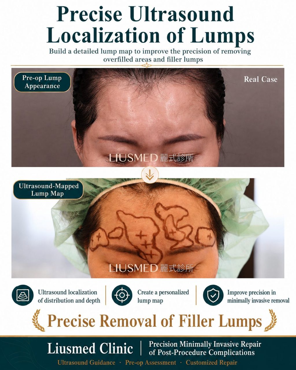
It's using ultrasound scanning to clearly visualize the lumps.
The lines in the photo aren't drawn randomly. They represent the areas, depths, and distribution confirmed by ultrasound, piece by piece—
A lump map specifically for her.
The biggest fear in filler lump revision has never been the size of the lump, but blindly dissecting or digging without knowing its exact location.
Especially with collagen stimulators, which often cause strong bodily reactions and are frequently encapsulated by layers of fibrous tissue. Relying solely on touch or experience can easily lead to unnecessary incisions, incomplete removal, or even damage to normal tissue.
Therefore, our sequence is: first, scan for precise localization, then mark, plan the removal path, and finally perform minimally invasive extraction.
We don't cut open the face to search for lumps. Instead, we visualize clearly first, then use the smallest possible entry point to treat only the areas that truly need attention.
She thought she would be left with five scars.
What I want to give her is not surgical scars, but the opportunity to restore her overfilled face, little by little.
Precise filler lump removal begins with pre-operative ultrasound scanning and localization.
Liusmed Clinic | Precise Minimally Invasive Post-Procedure Complication Revision (Real Case)
#FillerLumps #CollagenStimulators #OverfilledCorrection #UltrasoundGuided #LiusmedClinic